20939803
Description
Flashcards by Pooja Acharya, updated more than 1 year ago
|
|
Created by Pooja Acharya
about 4 years ago
|
|
| Question | Answer |
| Define pathogenesis | How a disease develops (the steps in the development of the disease) |
| A normal cell experiences stress. What outcomes may arise from this stress? | 1) the cell will adapt 2) if adaptation is non-viable, cell injury occurs. |
| What are the 4 types of cellular adaptation? | 1) hypertrophy 2) hyperplasia 3) atrophy 4) metaplasia |
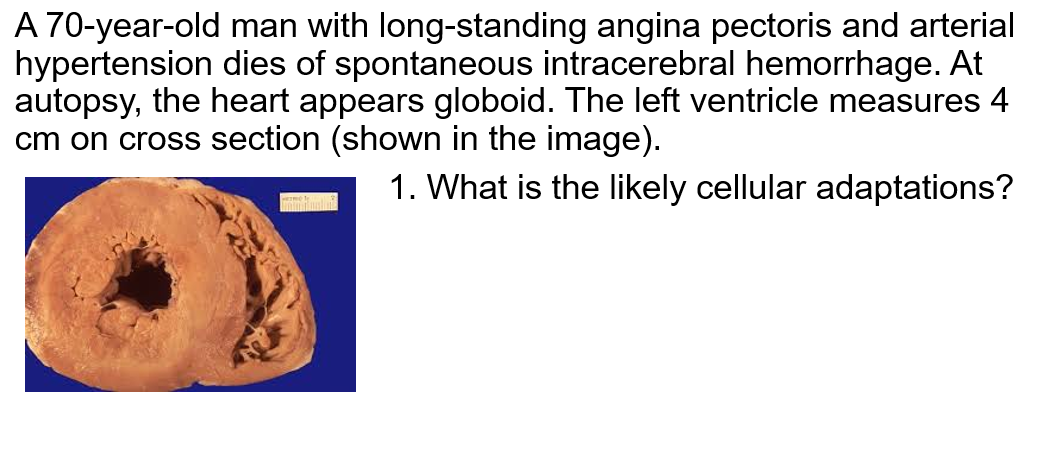
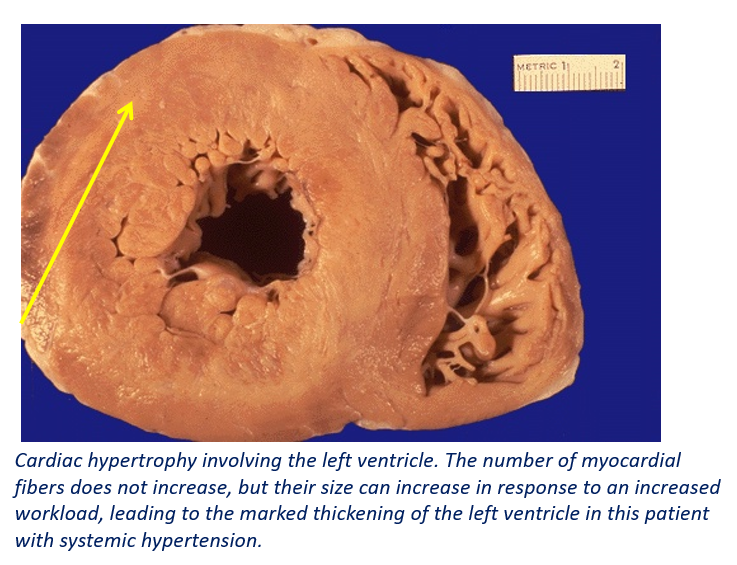
| Define Hypertrophy - What type of cells utlize this adaptive mechanism? - what occurs during this adaptation? | Hypertrophy is an increase in the size of cells, resulting in an increased organ size. no new cells arise - Occurs in permanent tissue i.e.with cells have limited capacity to divide (Nerves, Skeletal and cardiac muscle). - an increase in structural proteins and organelles also occurs. |
| What causes a cell to undergo hypertrophy? If giving examples, are they pathological or physiological? | 1) Growth factors or hormonal stimulation (e.g., pregnancy - uterine enlargement) (physiological) 2) increased functional demand - HTN (pathological) - Skeletal muscle in body builders (physiological) |
| Define Hyperplasia - what type of cells are capable of this? | An increase in the number of cells in an organ or tissue - only occurs in cells which synthesize DNA (NOT Nerve, Cardiac or Skeletal muscle) |
| Name 6 causes for atrophy | 1) decreased workload (disuse) 2) loss of innervation (denervation atrophy) 3) diminished blood supply 4) inadequate nutrition 5) loss of endocrine stimulation 6) aging (senile atrophy) |
| What are the 2 mechanisms of atrophy | 1) decreased protein synthesis (reduced metabolic activity) 2) increased protein degradation - autophagy - ubiquitin proteasome pathway |
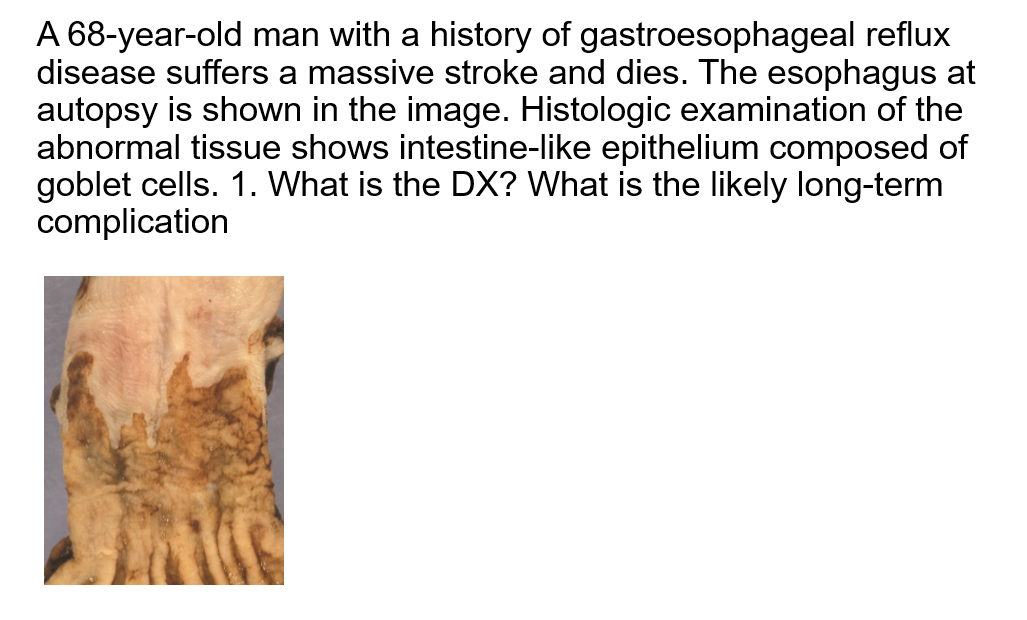
| Define metaplasia | Metaplasia is a reversible change in which one adult cell type is replaced by another in order to handle a given stress. |
| In chronic gastric reflux what adaptation is expected to occur? | Squamous epithelium turn columnar in this metaplastic change. (Barrott esophagus). |
| Cellular swelling is a morphology of reversible cell injury, what is the mechanism___________? | A failure of energy-dependent ion pumps in the plasma membrane. |
| What are the morphological features of reversible cell injury? | 1) plasma membrane alterations (blebbing, distortion of microvilli, loosening of intracellular attachements) 2) mitcohondrial changes (swelling) 3) ER dilation (detachment of ribosomes 4) Nuclear alterations (clumping of chromatin) 5) myelin figures (phospholipd masses from damaged cellular membrane) |
| What is the morphology of necrosis? | Pyknotisis --> karyorrhexis--.Karyolysis - breakdown of plasma membrane and organellar membranes - leakage and enzymatic digestion of cellular contents - myelin figures |
| What are the 6 patterns of tissue necrosis? | 1) coagulative necrosisthe most common) 2) liquefaction necrosis 3) caseous necrosis 4) Fat necrosis 5) fibrinoid necrosis - only observed histologically. 6) gangrenous necrosis |
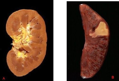
| Define coagulative necrosis. - what are key features of this form of necrosis? |
Coagulation is the most common form of necrosis
- denaturation of cytoplasmic proteins w/ preservation of framework of cells for several days (tissue integrety remains)
- characteristic of infarcts in all organs EXCEPT THE BRAIN
Image:
1 (binary/octet-stream)
|
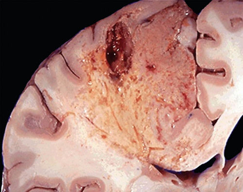
| Define liquefactive necrosis - what are key features of this necrotic form? - what structures undergo this form of necrosis? |
Necrotic area which is soft, and filled with fluid. The cellular framework is destroyed
- localized bacterial infections (abscesses) and found only in the brain
Image:
2 (binary/octet-stream)
|
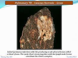
| What is caseous necrosis? What is a common disease that this necrosis occurs in? |
A soft, cottage cheese-like necrosis
- found in tuberculosis(Due to lipid rich mycolic acid of bacterial cell wall)
Image:
3 (binary/octet-stream)
|
|
Image:
4 (binary/octet-stream)
|
Image:
5 (binary/octet-stream)
|
|
Image:
6 (binary/octet-stream)
|
In Barrett esophagus, Squamous epithelium at the lower end of esophagus is replaced by Intestinal epithelium with goblet cells – intestinal metaplasia – a risk of developing adenocarcinoma of the esophagus |
|
Image:
7 (binary/octet-stream)
|
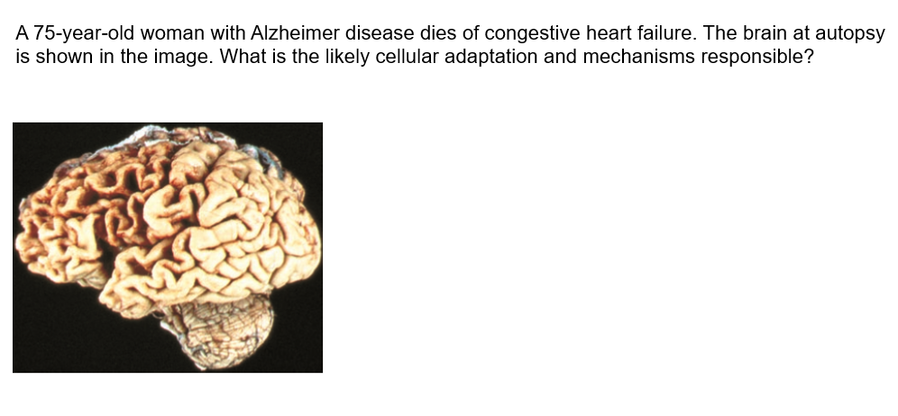
1. Atrophy:Note that loss of brain substance narrows the gyri and widens the sulci. The meninges have been stripped from the right half of each specimen to reveal the surface of the brain. 2. Autophagy, ubiquitin proteasomal degradation of proteins |
| Concentric hypertrophy of the Heart is seen in _____________ | Prolonged HTN, Aortic stenosis |
| Complication of endometrial hyperplasia__________ | Endometrial adenocarcinoma |
| Streak ovaries in Turner syndrome is an example of_____________ | Hypoplasia |
| Lipofuscin residual bodies in the cytoplasm would represent? _______ | Lipid peroxidation products resides in the lysosomes (Not pathological) |
| Cancer cachexia is due to an increased secretion of which cytokine___________ | TNF |
| Mechanism of Metaplasia_________ | Reprogram of stem cells to withstand noxious stimuli |
| What is fat necrosis? - what mechanism allows for fat necrosis to occur? | - focal areas of fat destruction - enzymes (macrophage or pancreatic cells) degrade FAs thath complex with calcium to create soaps, grossly white and chalky |
| What is fibrinoid necrosis? |
Immune complexes, together with fibrin leaking out of a blood vessel (only seen histologically).
Image:
8 (binary/octet-stream)
|
| What are the principal targets and biochemical mechanisms of cell injury? (4) | 1) mitochondria and their ability to generate ATP and ROS under pathologic conditions 2) disturbances in calcium homeostasis 3) damage to cellular membranes (plasma and lysosomal) 4) damage to DNA and misfolding of proteins. |
| What consequences occur when cell injury leads to an increased calcium influx? (5 | phosopholipase is activated, which damages the cell membranes phospholipds 2) protease activation disrupts proteins 3) endonuclease activation breaks down the nuclease 4) activation of ATPase results in decreased ATP concentrations 5) mitochondrial permeability transition: opens up pores increasing Ca permeability, disurpint the Electro-potential. |
| How do phagosomes create free radicals, what enzyme helps with this process and what purpose does this mechanism serve? | phagosomes use phagocyte oxidase to convert NADPH into H2O2, which can then be converted into bleach - used to destroy microbial agents. |
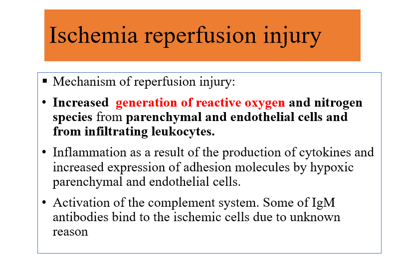
| What is ischemia reperfusion injury? Explain pathophysiological mechanisms responsible. |
Blood flow to ischemic tissue can be restored, which can promote recovery of cells if they are reversibly injured but may paradoxically exacerbate the injury and result in cell death(Following tissue plasminogen activator)
Image:
9 (binary/octet-stream)
|
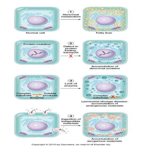
| What are the for pathologies of intracelluar accumulations |
Image:
10 (binary/octet-stream)
|
| What are the two typesof lipid accumulations that occur intracellularly? What are their etiologies? | 1) triglyceride accumulation (steatosis) - Etiologies: - toxins, protein malnutrition, DM, obesity or anoxia 2) cholesterol and cholesterol esters (atherosclerosis) |
| Give 2 examples of endogenous pigments | Lipofuscin & melanin |
| Proapoptotic factors __________ | BAX, BAD,BIM |
| Anti-apoptotic factors | BCL-2 |
| Killing of virus infected cells by cytotoxic T-Cells_______________ | Perforins and granzymes |
| Accumulation of misfolded proteins due to ER stress is an example of________________ | Cystic fibrosis, Huntingtons, Alzhiemer, A1 antitrypsin def |
| Ink dot nucleus(pyknosis) and eosinophilic cytoplasm with intact cell membrane on light microscopy, this feature suggest_______ | Apoptotic body |
| Mutations of bcl-2 can lead to ___________ | Follicular lymphoma |
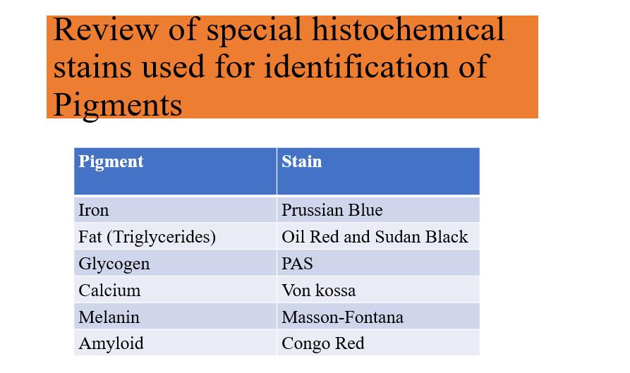
| Mention special stains used to diagnose various intracellular accumulations> |
Image:
11 (binary/octet-stream)
|
Want to create your own Flashcards for free with GoConqr? Learn more.