30240986
Description
Flashcards by aarthi kannan, updated more than 1 year ago
|
|
Created by aarthi kannan
over 4 years ago
|
|
| Question | Answer |
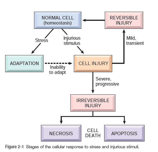
| What are the stages of cellular response to stress and injurious stimuli? | |
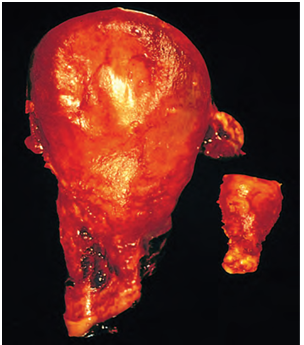
| What change has occurred in this organ? Explain the mechanism ? Left – specimen of uterus removed after postpartum hemorrhage Right – specimen of uterus from normal non gravid person | Answer: Physiologic hypertrophy of the uterus during pregnancy. |
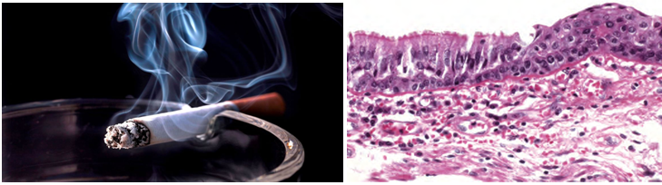
| Connect the images and explain the change seen in the bronchial mucosa. What is the underlying mechanism? | The pathological change seen here is metaplasia. Metaplasia is a reversible change in which one differentiated cell type (epithelial or mesenchymal) is replaced by another cell type. Metaplasia does not result from a change in the phenotype of an already differentiated cell type; instead, it is the result of reprogramming of stem cells that are known to exist in normal tissues, or of undifferentiated mesenchymal cells present in connective tissue. |
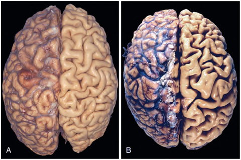
| A. Normal brain of young adult B. Brain in 85-year-old man with atherosclerosis of cerebral arteries What change has occurred here? What are the causes for this change? | The old man has cerebral atrophy due to reduced blood supply from atherosclerotic blood vessels. There is widening of sulci and narrowing of gyri when compared to the normal brain. Atrophy results from decreased protein synthesis and increased protein degradation in cells. The causes of Atrophy include: 1. Decreased workload ( atrophy of disuse) 2. Loss of innervations ( denervation atrophy) 3. Diminished blood supply 4. Inadequate nutrition. 5. Loss of endocrine stimulation 6. Pressure |
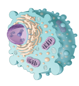
| What are the changes associated with reversible cell injury? | 1. Decreased ATP levels 2. Ion and fluid imbalance 3. Cellular swelling 4. Decreased pH 5. Fatty change 6. Swelling of mitochondria 7.Ribosomal detachment from ER |
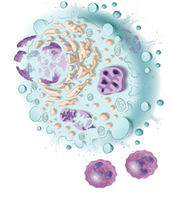
| What are the changes associated with irreversible cell injury? | 1. Amorphous densities in mitochondria 2. Severe membrane damage 3. Lysosomal rupture 4. Extensive DNA damage |
| What are the intracellular mechanisms of cell injury? | 1. Depletion of ATP 2. Mitochondrial damage 3. Loss of calcium homeostasis with influx of calcium 4. Oxidative stress (accumulation of O2 free radicals) 5. Defects in membrane permeability 6. Damage to DNA & proteins |
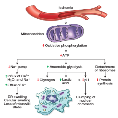
| Describe the functional and morphological consequences of decreased intracellular adenosine triphosphate (ATP) during cell injury | |
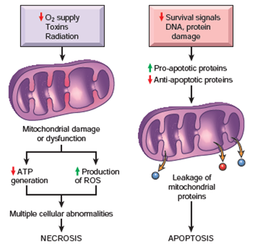
| Describe the role of mitochondria in cell injury and cell death | |
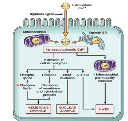
| Describe schematically the role of increased cytosolic calcium in cell injury | |
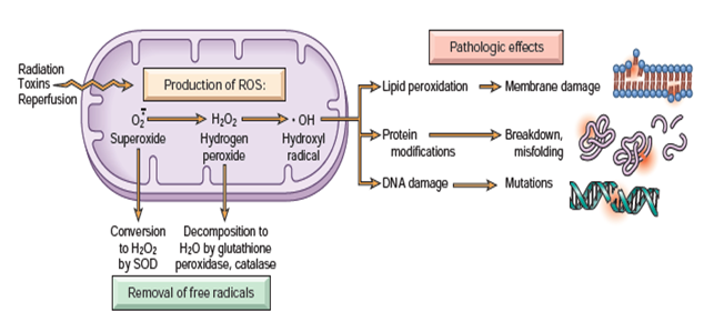
| Diagrammatic representation of the mechanism of cell injury by free radicals | |
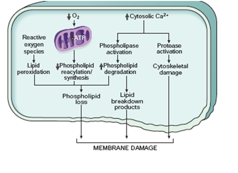
| Describe the mechanisms of membrane damage in cell injury | |
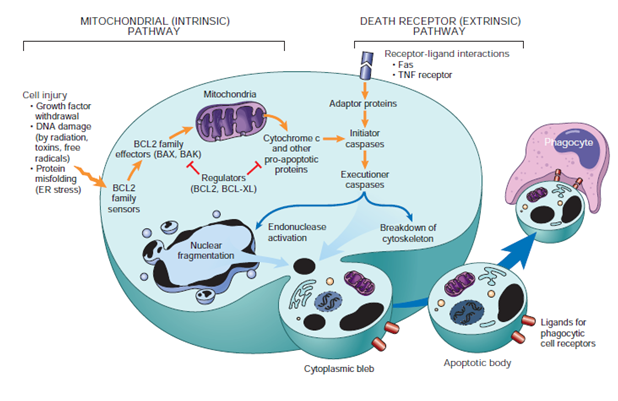
| Schematically describe the mechanisms of apoptosis through the intrinsic and extrinsic pathway | |
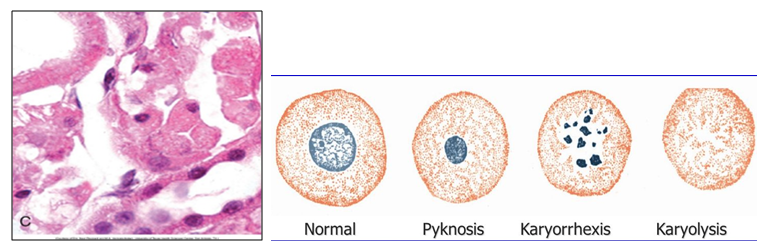
| Enlist the morphological changes in necrosis. | • Increased eosinophilia • Glassy homogenous – loss of glycogen • Vacuolated, moth-eaten cytoplasm • Calcification of dead cells • Myelin figures – phospholipid precipitates • Fluffy denatured protein • Mitochondrial dense bodies (calcium)* • Discontinuities in plasma and organelle membranes • Nuclear changes of pyknosis, karyorrhexis, karyolysis |
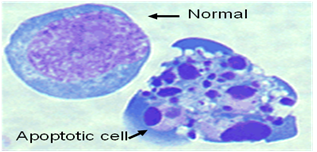
| What are the morphological changes in apoptosis? | 1. Cell shrinkage 2. Chromatin Condensation 3. Intact cell membrane until late 4. Formation of cytoplasmic blebs and apoptotic bodies 5. No inflammation 6. Phagocytosis of apoptotic bodies |
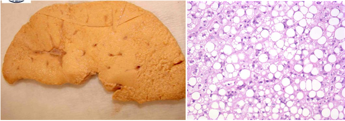
| Explain the changes that have taken place in the Liver? | The image shows fatty liver (steatosis). When delivery of free fatty acids to the liver is increased, as in diabetes or when intrahepatic lipid metabolism is disturbed, as in alcoholism, triglycerides accumulate in liver cells |
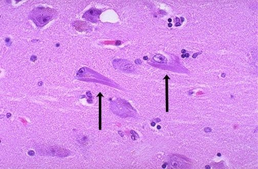
| Identify this pathological accumulation seen in the hippocampal area of the Brain from an 85-year-old male | This is neurofibrillary tangle found in the brain in Alzheimer's disease and it contains neurofilaments and other proteins. Accumulations of keratin filaments and neurofilaments are associated with certain types of cell injury. |
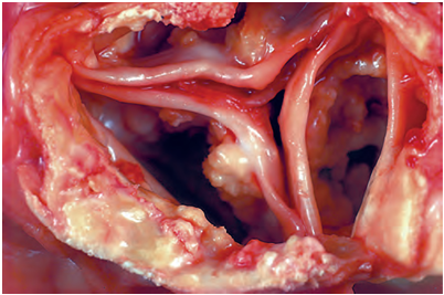
| Describe this pathological change in cardiac valves. What special stains is used to recognize this change? | This is Dystrophic calcification of the aortic valve. The valve is markedly narrowed (stenosis). The semilunar cusps are thickened and fibrotic, and behind each cusp are irregular masses of piled-up dystrophic Calcification. In H & E sections, they appear basophilic amorphous & granular masses. The calcium can be demonstrated by Von Kossa stain. |
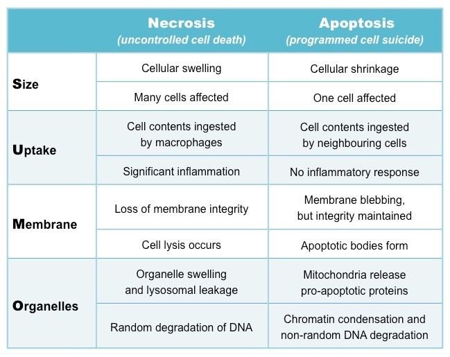
| List the differences between Necrosis and Apoptosis | |
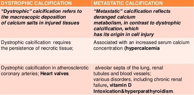
| What are the differences between dystrophic and metastatic calcification? |
Want to create your own Flashcards for free with GoConqr? Learn more.