
Flashcards
Physiology semester 2
Cards in this deck (85)
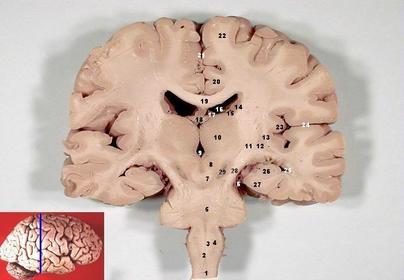
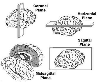
- 1Define: Frontal sectionA slice through the brain parallel to the forehead
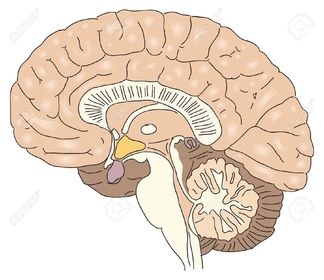
- 2Define : Sagittal sectionA slice through the brain parallel to the neuraxis and perpendicular to the ground
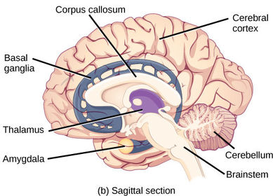
- 3Define : Cross sectionWith respect to the CNS, a slice taken at right angle to the neuraxis
- 4Define : Medial and LateralMedial = Toward the middle of the body away from the side
Lateral = Toward the side away from the middle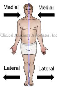
- 5Define : Horizontal sectionA slice through the brain parallel to the ground
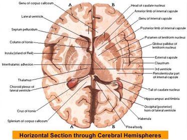
- 6Define : Midsagittal planeThe plane through the neuraxis perpendicular to the ground, divides the brain into two symmetrical halves
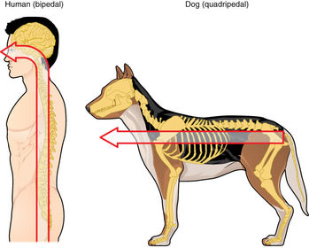
- 7Define : NeuraxisAn imaginary line drawn through the center of the length of the CNS, from the bottom of the spinal cord to the front of forebrain
- 8Define the terms Ipsilateral and contralateralIpsilateral = Located on the same side of the body
Contralateral = Located on the opposite side of the body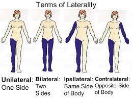
- 9Define : VentralVentral = 'toward the belly' in a direction perpendicular to neuraxis toward the bottom of the skull or front surface of the body
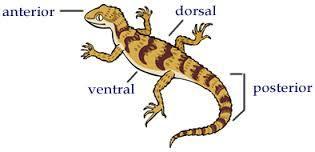
- 10Define : DorsalDorsal = 'toward the back' in a direction perpendicular to neuraxis towards top of head or the back
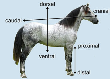
- 11Define : Rostral and CaudalRostral = 'toward the beak' with respect to the CNS, in a direction along the neuraxis towards front of the face
Caudal = 'toward the tail', in a direction along neuraxis away from the front of the face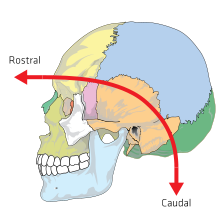
- 12Define : Anterior and PosteriorAnterior = near of toward the head
Posterior = near or toward the tail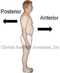
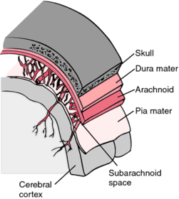
- 13Define : Dura materDura mater = outer later of the meninges, it's thick, tough and flexible but un-stretchable
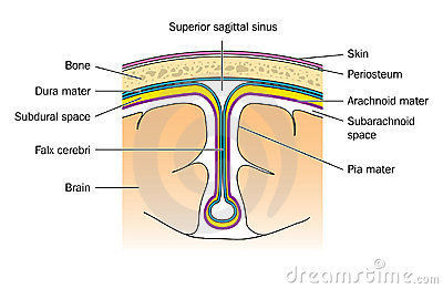
- 14What are the 3 layers of tissue that encase the CNS?Layers of meninges;
1. Dura mater
2. Arachnoid membane
3. Pia mater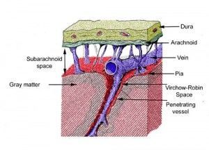
- 15Define : Arachnoid membraneThe arachnoid membrane is the middle layer of the meninges. It's soft, spongy and lies beneath the dura mater
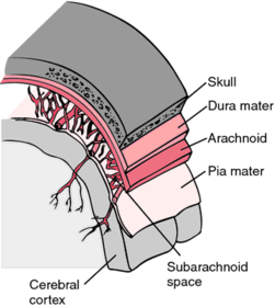
- 16Define : Pia materThe pia mater is a layer of the meninges which clings to the surface of the brain. It is thin and delicate
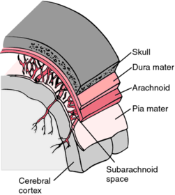
- 17What is the subarachnoid space?it is a fluid filled space , (cerebrospinal fluid ) that cushions the brain, located between the arachnoid membrane and pia mater.
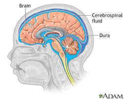
- 18What is cerebral spinal fluid?Clear fluid similar to blood plasma that fills the ventricular system of brain and sub-arachnoid space surrounding brain and spinal cord
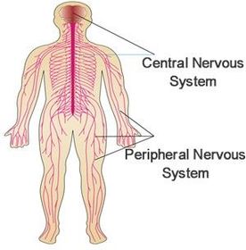
- 19What is the central nervous system made up of?- The brain - mass of neurons, glia and other supporting cells. Most protected organ, encased in a tough bony skulls and floating in cerebrospinal fluid. receives 20% of the blood flow from heart and is chemically protected by blood brain barrier
- Spinal cord - 20What is the peripheral nervous system covered by?2 layers of the meninges -
Dura mater and Pia mater fuse and form a sheath that covers the spinal and cranial nerves and peripheral ganglia. - 21What is the peripheral nervous system made up of ?Spinal and cranial nerves and peripheral ganglia
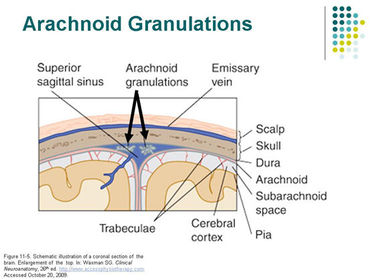
- 22Where is cerebrospinal fluid produced and how is it transported?Produced in the choroid plexus of the lateral, third and fourth ventricles. It flows from the two lateral ventricles into the third ventricle, through the cerebral aqueduct into the fouth ventricle , then into the subarachnoid space, and finally back into the blood supply through the arachnoid granulations
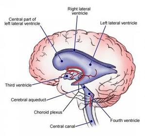
- 23What happens when the flow of cerebrospinal fluid is blocked by a tumor or other obstruction?It results in hydrocephalus: enlargement of the ventricles and subsequent brain damage
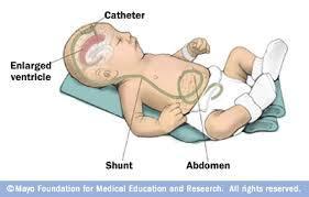
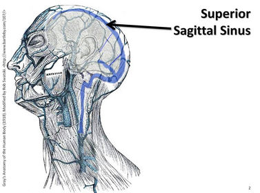
- 24What is the superior sagittal sinus?A venous sinus located in the midline jusr dorsal to the corpus callosum, between the two cerebral hemispheres
- 25What is arachnoid granulation?small projections of the arachnoid membrane through the dura mater into the superior sagittal sinus; CSF flows through them to be reabsorbed into the blood supply
- 26What is the cerebral cortex?The outermost layer of gray matter of the cerebral hemispheres.
Consists mostly of glia & cell bodies, dendrites and interconnecting axons of neurons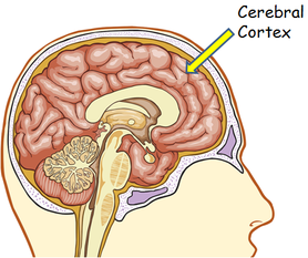
- 27What are progenitor cells?Cells of the ventricular zone that divide and give rise to cells of the central nervous system.
- 28What is neurogenesis?Production of new neurons through the division of neural stem cells. Occurs in hippocampus and olfactory bulb and appears to play a role in learning.
- 29What is apoptosis?Death of a cell caused by a chemical signal that activates a genetic mechanism inside the cell.
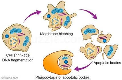
- 30What is symmetrical and asymmetrical division?Symmetrical division = Division of a progenitor cell that gives rise to 2 identical progenitor cells and increases size of VZ and hence brain develops from it.
Asymmetrical division = Division of progenitor cell that gives rise to another progenitor and a neuron which migrates away from VZ toward final resting place in the brain. - 31What is the ventricular zone?A layer of cells that line to the inside of the neural tube; contains progenitor cells that divide and give rise to cells of the CNS.
- 32What is radial glia?Special glia with fibers that grow radially outward from the VZ to the surface of the cortex; provide guidance for neurons migrating outward during brain development.
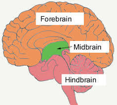
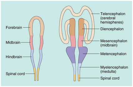
- 33What is the forebrain?The most rostral of the 3 major divisions of the brain and includes telencephalon and diencephalon.
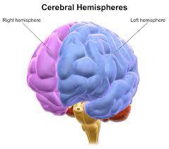
- 34What is the cerebral hemisphere?One of the two major portions of the forebrain covered by cerebral cortex
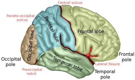
- 35Define : Lateral fissureA fissure that separates the temporal lobe from the overlying frontal and parietal lobes.
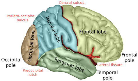
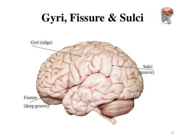
- 36Define : SulcusA groove in the surface of the cerebral hemisphere, smaller than a fissure
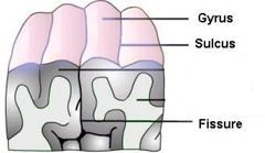
- 37Define : FissureA major groove in the surface of the brain
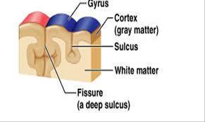
- 38Define : GyrusA convolution of the cortex of the cerebral hemispheres separated by sulci or fissures
- 39Define : central sulcusSulcus that separates the frontal lobe from the parietal lobe
- 40Why is it called white matter?Due to the large concentration of myelin which has a white opaque appearance
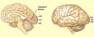
- 41What is the calcarine fissure?A fissure located in the occipital lobe on the medial surface of the brain, most of the primary visual cortex is located along its upper and lower banks.
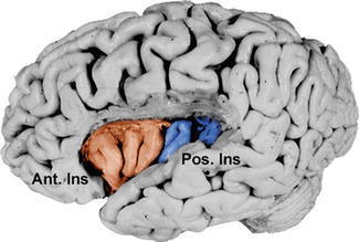
- 42What is the insular cortex?A sunken region of the cerebral cortex that is normally covered by the rostral superior temporal lobe and caudal inferior frontal lobe.
- 43Describe the telencephalon...Most of 2 symmetrical cerebral hemispheres that make up cerebrum. Cerebral hemispheres covered by cerebral cortex and contain limbic system and basal ganglia in subcortical regions
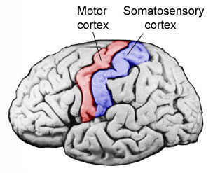
- 44Where is the primary somatosensory cortex?In the region of the anterior parietal lobe whose primary input is form the somatosensory system.
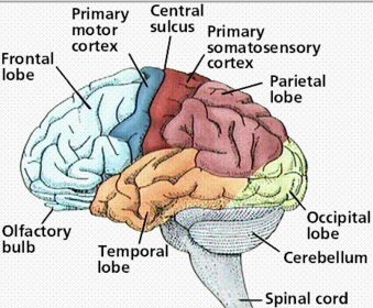
- 45Where is the primary visual cortex?The region of the posterior occipital lobe whose primary input is from the visual system
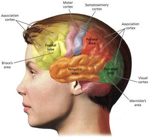
- 46Where is the primary motor cortex?Region of the posterior frontal lobe that contains neurons that control movements of skeletal muscles
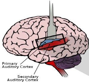
- 47Where is the primary auditory cortex?The region of the superior temporal lobe whose primary input is from the auditory system.
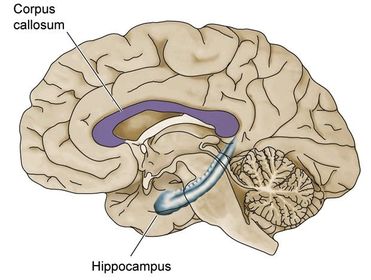
- 48Define : Corpus callosumA large bundle of axons that interconnects corresponding regions of the association cortex on each side of the brain ( left and right hemisphere
- 49Define : Cingulate gyrusA strip of limbic cortex lying along the later walls of the groove seperating the cerebral hemispheres, just above the corpus callosum.
- 50Define : Limbic systemA group of brain regions including the anterior thalamic nuclei, amygdala, hippocampus, limbic cortex, and parts of the hypothalamus, as well as their interconnecting fiber bundles. Involved in emotion, motivation and learning.
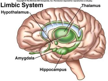
- 51Define : HippocampusA forebrain structure of the temporal lobe, constituting an important part of the limbic system; includes the hippocampus proper, dentate gyrus and subiculum.
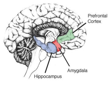
- 52Define : AmygdalaA structure in the interior if the rostral temporal lobe, containing a set of nuclei; part of the limbic system.
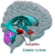
- 53Define : Fornix Mammilary bodiesFornix = A fiber bundle that connects the hippocampus with other parts of the brain, including the mammillary bodies of the hypothalamus; part of the limbic system.
Mammillary bodies = A protrusion of the bottom of the brain at the posterior end of the hypothalamus, containing some hypothalamic nuclei; part of the limbic system - 54Define : Basal gangliaA group of subcortical nuclei in the telencephalon, the caudate nucleus, the globe pallidus, and the putamen; important parts pf the motor system. Participates in the control of movement
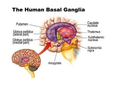
- 55Define : DiencephalonA region of the forebrain surrounding the third ventricle; includes the thalamus and hypothalamus
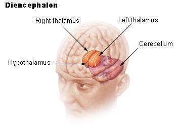
- 56Define : thalamusThe largest portion of the diencephalon, located above the hypothalamus; contains nuclei that project information to specific regions of the cerebral cortex and receive information from it.
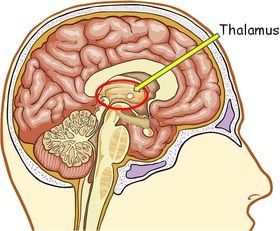
- 57Define : lateral geniculate nucleus Medial geniculate nucleus Ventrolateral nucleuslateral geniculate nucleus = A group of cell bodies within the lateral geniculate body of the thalamus that received fibers from the retina and projects fibers to the primary visual cortex
Medial geniculate nucleus = A group of cell bodies within the medial geniculate body of the thalamus; receives fibers from the auditory system and projects fibers to the primary auditory cortex
Ventrolateral nucleus - A nucleus of the thalamus that received inpites from the cerebellum and sends axons to the primary motor cortex - 58Define : HypothalamusThe group of nuclei of the diencephalon situated beneath the thalamus; involved in regulation of the autonomic nervous system, control of the anterior and posterior pituitary glands, and integration of species-typical behaviours
- 59Define : Anterior pituitary gland and posterior pituitary glandThe anterior part of the pituitary gland; an endocrine gland whose secretions are controlled by the hypothalamic hormones
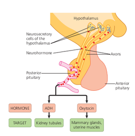
Posterior pituitary gland = an endocrine gland that contains hormone-secreting terminal buttons of axons whose cell bodies lie within the hypothalamus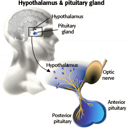
- 60Define : Neurosecretory cellA neuron that secretes a hormone of hormone-like substance
- 61Define : Midbrain / mesencephalonA region of the brain that surrounds the cerebral aqueduct ; includes the tectum and the tegmentum
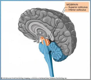
- 62What are the 2 main structures found in the tectum?Superior colliculi = protrusions on top of the mid brain - part of the visual system
Inferior colliculi = Protrusions on the top of the mid brain - part of the auditory system - 63Define : Tegmentum?The ventral part of the midbrain; includes the periaqueductal gray matter, reticular formation, red necleus and substantia nigra.
- 64What is the red nucleus and where is it found?Found in Tegmentum
A large nucleus of the midbrain that recives input from the cerebellum and motor cortex and sends axons to motor neurons in the spinal cord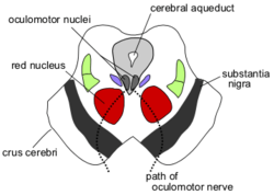
- 65Define : HindbrainThe most caudal of the 3 major divisions of the brain, it surrounds the fourth ventricle and includes the metencephalon and myelencephalon
- 66Define : Cerebellum"little brain" a major part of the brain located dorsal to the pons, containing two cerebellar hemispheres, covered with the cerebellar cortex; an important component of the motor system. Plays important role in integrating and coordinating movements.
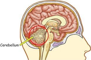
- 67Define : deep cerebellar nuclei and cerebellar peduncleDeep cerebellar nuclei = Nuclei located within the cerebellar hemispheres; receive projections from the cerebellar cortex and send projections out of the cerebellum to other parts of the brain
Cerebellar peduncle = one of three bundles of axons that attach each cerebellar hemisphere to the dorsal pons - 68Define : PonsThe region of the metencephalon rostral to the medulla, caudal to the midbrain and ventral to the cerebellum. The pons contains in its core, a portion of the reticular formation, including some nuclei that appear to be important in sleep and arousal. It also contains a large nucleus that relays information from the cerebral cortex to the cerebellum.
- 69Define : Medulla OblongataThe most caudal potion of the brain; located in the myelencephalon, immediately rostral to the spinal cord. The medulla contains part of the reticular formation, including nuclei that control vital functions such as regulation of the cardiovascular system, respiration and skeletal muscle tonus. Also may be involved in sleep and arousal
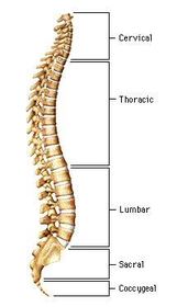
- 70What is the principle function of the spinal cord?To distribute motor fibers to the effector organs of the body (glands and muscles) and to collect somatosensory information to be passed on to the brain.
- 71Define : spinal rootA bundle of axons surrounded by connective tissue that occurs in pairs, which fuse and form a spinal nerve.
- 72Define : Caudal blockThe anaesthesia and paralysis of the lower part of the body produced by injection of local anaesthetic into cerebrospinal fluid surrounding the cauda equina
- 73Define : dorsal rootThe spinal root that contains incoming sensory fibers (afferent)
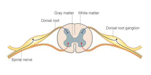
- 74Define : Ventral rootThe spinal root that contains outgoing (efferent) motor fibers.
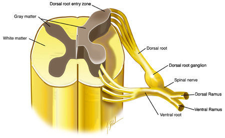
- 75How does the CNS form? Describe the stagesDuring the first phase of brain development, symmetrical division of the progenitor cells of the vz and svz, which lines the neural tube, increase in size. During the second phase, asymmetrical division of these cells gives rise to neurons, which migrate up the fibres or radial glial cells to their final resting place. There, neurons develop dendrites and axons and establish synaptic connections with other neurons. Later, neurons that fail to develop a sufficient number of synpatic connections are killed through apoptosis. Although the basic development on CNS is genetically controlled, sensory stimulation plays a role in refining the details. In addition, the neural circuitry of even a full grown mature brain can be modified through experience.
- 76Define : Vagus nerveThe largest of the cranial nerves, conveying efferent fibers of the parasympathetic division of the ANS to organs of the thoracic and abdominal cavities
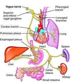
- 77Define : Dorsal root ganglionA nodule on a dorsal root that contains cell bodies of afferent spinal nerve neurons
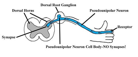
- 78What is the difference between efferent and afferent axons?Efferent = An axon directed AWAY from CNS, conveying motor commands to muscles and glands
Afferent - An axon directed TOWARDS the CNS, conveying sensory information. - 79Define : Cranial nerveA peripheral never attached directly to the brain
- 80What do all of the axons in the dorsal root convey?Somatosensory information
- 81Define : Somatic nervous systemThe part of the peripheral nervous system that controls the movement of skeletal muscles or transmits somatosensory information to the CNS+
- 82Define : Autonomic nervous system, Sympathetic and parasympathetic divisionANS = The portion of the peripheral nervous system that controls the bodys vegetative functions
Sympathetic division = Portion of ANS that controls functions that accompany arousal and expenditure of energy ( such as increased heart rate)
Parasympathetic division = Portion of ANS that controls functions that occur during a relaxed state (such as decreased heart rate). - 83Define : Sympathetic gangliaNodules that contain synapses between preganglionic and postganglionic neurons of the sympathetic nervous system.
- 84Define : postganglionic neuron and preganglionic neuronPreganglionic neuron = The efferent neuron of the ANS whose cell body is located in a cranial nerve nucleus or in the intermediate horn of the spinal grey matter and whose terminal buttons synpase upon postganglionic neurons in the autonomic ganglia
Postganglionic neuron= Neurons of the ANS that form synapses directly with their target organ - 85Define : Adrenal medullaThe adrenal medulla (Latin: medulla glandulae suprarenalis) is part of the adrenal gland. It is located at the center of the gland, being surrounded by the adrenal cortex. It is the innermost part of the adrenal gland, consisting of cells that secrete epinephrine (adrenaline), norepinephrine (noradrenaline). It is controlled by axons of the sympathetic nervous system.
Information
- Description
- Degree Psychology (Physiology) Flashcards on Physiology semester 2 , created by Molly Macgregor on 13/04/2016.
- Created by
- Molly Macgregor
- Views
- 5
- Rating