6301333

| Question | Answer |
| 3.8 The control of gene expression | N/A |
| 3.8.1 Alteration of the sequence of bases in DNA can alter the structure of proteins | N/A |
| When might gene mutations arise? | During DNA replication |
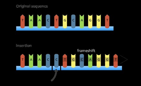
| What is an addition mutation? | Additional base inserted into the strand, causing frame shift to the right - can cause every protein after the addition to be different |
| Diagram of an addition mutation | |
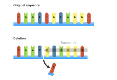
| What is a deletion mutation? | Base deleted from the strand, causing frame shift to the left - can cause every protein after the addition to be different |
| Diagram of an deletion mutation | |
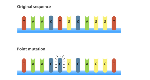
| What is a substitution mutation? | Base from the strand replaced by another - can cause the protein coded for by this triplet to be different |
| Diagram of a substitution mutation | |
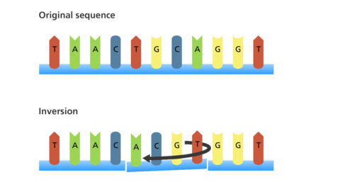
| What is an inversion mutation? | Section of the strand is inverted horizontally - can cause protein coded for by any of the codons effected to be changed (depending on the number of bases in the section, whether it is palindromic etc) |
| Diagram of an inversion mutation | |
| What is a duplication mutation? | Base duplicated and inserted into the strand beside the original, causing frame shift to the right - can cause every protein after the addition to be different |
| Diagram of a duplication mutation | |
| What is a translocation mutation? | Section of DNA strand is separated from one chromosome and inserted into another, causing significant frame shift (addition of multiple bases) - can cause every protein after the addition to be different |
| Diagram of a translocation mutation | |
| What can mutations result in? | Different amino acid sequence in the encoded polypeptide |
| What impact do mutagenic agents have? | Increase the rate of mutations |
| What are some examples of mutagenic agents? | - High energy ionising radiation - can disrupt the structure of DNA - Chemicals e.g. nitrous dioxide may alter DNA structure or interfere with transcription |
| 3.8.2 Gene expression is controlled by a number of features | N/A |
| 3.8.2.1 Most of a cell’s DNA is not translated | N/A |
| What are totipotent cells? | Cells able to divide and produce any kind of cell in an organism - found in early mammal embryos for a short time |
| How does cell specialisation occur? | Totipotent cells translate only part of their DNA |
| What are pluripotent cells? | Able to differentiate into almost any type of cell - found in embryos |
| What are multipotent cells? | Can differentiate into a limited number of specialised cells, usually develop into cells of a particular type e.g. stem cells in bone marrow can produce any type of blood cell - found in adult mammals |
| What are unipotent cells? | Can only differentiate into a single type of cell, derived from multipotent stem cells - found in adult mammals |
| How could pluripotent stem cells be used in medicine? | Divide in unlimited numbers - repair damaged tissues e.g. burned skin, Parkinson's - reverse effects of paralysis by growing new nerve cells - cure type I diabetes by transplanting insulin-producing cells |
| What are Induced Pluripotent Stem Cells (iPS cells)? | Unipotent stem cells genetically altered to be pluripotent, done by modifying genes and transcription factors - able to self-replicate for limitless supply - could replace embryonic stem cells (not self replicating) in research, thus overcoming ethical concerns |
| What are some arguments for the use of embryonic stem cells in research? | - Embryo has no status, is not considered alive/having valuable life - There is a moral obligation to aid sufferers - Research can help to develop cures of chronic conditions - Could result in treatments for previously untreatable conditions |
| What are some arguments against the use of embryonic stem cells in research? | - Requires the killing of a potential life (blastocyst must be destroyed to acquire stem cells) |
| 3.8.2.2 Regulation of transcription and translation | N/A |
| What are transcriptional factors? | Factors that cause stimulation or inhibition of transcription by their movement from cytoplasm into the nucleus |
| What is the role of oestrogen in the stimulation of transcription? | - Oestrogen diffuses through cell membrane into cytoplasm - Binds to a receptor, ER-alpha (transcription factor held in protein complex to inhibit it) - ER-alpha receptor changes shape - ER-alpha oestrogen receptor leaves protein complex and attaches to promoter region of the target gene - Other co-factors are attracted and also bind to the promoter region - RNA polymerase begins transcription of the target gene |
| What is epigenetics? | The study of changes in organisms caused by modification of gene expression rather than alteration of the genetic code itself |
| How can epigenetics alter gene expression? | Make heritable changes to gene expression in eukaryotic cells, without alterations to DNA base structure |
| What is the epigenome? | Chemical tags that form the second layer of the histone and determine their shape - keeps inactive genes tightly packed and ensures they can't be read - EPIGENETIC SILENCING - flexible, able to adapt to environment changes |
| What is the effect of condensation of the DNA-histone complex? | DNA more tightly condensed (coiled) around histones - means that part of the DNA can't be transcribed as it is inaccessable to transcription factors |
| In what ways can gene expression be altered? | Inhibition of transcription by: - decreased actylation of associated histones - increased methylation of DNA |
| How can decreased acetylation of associated histones affect gene expression? | Acetylation - attachment of acetyl group onto DNA, occurring constantly - acetyl group has negative charge, thus rate of acetylation decreasing increases positive charge of histone group - increased positivity attracts negative phosphate group on DNA, causing greater condensation and thus inhibiting transcription |
| How can increased methylation of DNA affect gene expression? | Methylation - Addition of methyl group (CH3) to DNA molecule, binds to cytosine base - Inhibits transcription of genes in two ways: - Prevents binding of transcriptional factors to DNA - attracts proteins that condense the DNA-histone complex by inducing de-acetylation of the histones, making part of the DNA inaccessable to transcription factors |
| How can epigenetics cause disease? | Increased methylation causes genes to be EPIGENETICALLY SILENCED - linked to cancer. Cancer cells have high methylation - Certain genes - TP53 gene codes for a protein that regulates cell division |
| How can epigenetics be used in treatment of disease? | Counteracting methylation could reactivate genes by inhibiting enzyme that causes methylation - treatment of cancer in this way would have to be very precise to avoid causing cancer in healthy cells - could also be used to make diagnostic cells - detection of methylation or low acetylation can be used to detect and locate cancerous cells |
| What environmental factors can influence epigenetics? | - Diet and stress can alter epigenome - Accumulation of lifestyle impacts |
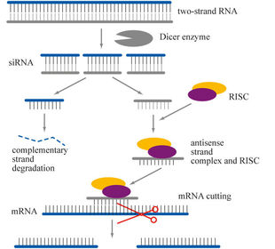
| Diagram of RNA interference | |
| What are the stages of RNA interference? | - enzyme cuts large double-stranded molecules of RNA into smaller sections (siRNA) - one of the siRNA strands combines with an enzyme - siRNA molecule guides the enzyme to a mRNA molecule by pairing up its bases to the complementary ones on a section of the mRNA - once in position, the enzyme cuts the mRNA into smaller sections - mRNA is no longer capable of being translated, thus this section is inhibitied |
| 3.8.2.3 Gene expression and cancer | N/A |
| What is a benign tumour? | Non-cancerous tumour |
| What are the characteristics of a benign tumour? | - Can grow to large size - Grow slowly - Cell nucleus has a relatively normal appearance - Cells are often well differentiated - Cells produce adhesion molecules that make them stick together and thus remain within the tissue in which they reside = primary tumours - Tumours surrounded by a capsule of dense tissue so remain compact structures - Much less likely to be life threatening but can disrupt functioning of a vital organ - Tend to have localised effects on the body - usually removed by surgery alone - Rarely reoccur after treatment |
| What is a malignant tumour? | Cancerous tumour |
| What are the characteristics of a malignant tumour? | - Can grow to a large size - Grow very rapidly - Cell nucleus often larger and darker due to abundance of DNA - Cells become de-differentiated - Cells do not produce adhesion molecules and so they spread to other body regions (metastasis), forming secondary tumours - Tumours are surrounded by a capsule and so can grow finger-like projections into the surrounding tissue - More likely to be life-threatening, abnormal tumour tissue replaces normal tissue - Often have systemic effects such as weight loss and fatigue - Removal usually involves radiotherapy and/or chemotherapy, as well as surgery - More frequently reoccur after treatment |
| What is the role of tumour-suppressor genes in regulating cell division? | Inherited in a pair - Slow cell division - Repair mistakes in DNA - Programme cell death (apoptosis) |
| How can tumour supressor genes be involved in development of tumours? | If mutated (both inactive): - inactivated, no longer inhibiting cell division & unable to repair DNA - Most mutated cells die, but some can survive to replicate freely. They can also move around the body due to lacking adhesion proteins - most failure of TS genes is acquired not inherited |
| What is the role of proto-oncogenes genes in stimulating cell division? | - Stimulate cell division when growth factors attach to a protein receptor on the cell-surface membrane |
| How can proto-oncogenes genes be involved in development of tumours? | - mutation into an oncogene can cause permanent activation, thus cell will be always stimulated to divide for two reasons: - receptor protein on the cell surface membrane can be permanently activated, so that cell division is always stimulated even in the absence of growth factors - oncogene may code for a growth factor that is produced in excessive amounts, stimulating cell division excessively |
| How can increased concentration of oestrogen increase the chance of breast cancer? | - Oestrogen is a transcription factor that triggers transcription, and with increased oestrogen (and therefore more transcription) more proteins will be synthesised - Some proteins are used to stimulate cell division and be receptors for stimulation on cell-surface membrane - Greater stimulation of cell division increases chance of cancer if TS genes inactive |
| 3.8.3 Using genome projects | N/A |
| What are sequencing projects? | Determining the sequence of all DNA in an organism |
| What are some methods of gene sequencing? | Whole-genome shotgun sequencing - DNA cut into small sections and then aligned to overlapping segments to assemble the entire genome using algorithms - rapid sequencing |
| What is the proteome? | All the proteins produced by the genome in a cell at a given time under specific conditions |
| What are the advantages of sequencing the genome of simple organisms? | - most prokaryotes have one circular DNA piece not associated with histones - no non-coding DNA portions |
| What are the possible applications of information gained from sequencing the genome of simple organisms? | - study organisms that endure extreme or toxic conditions for use in cleaning up pollutants/manufacturing biofuels - identification of potential antigens for use in vaccine production e.g. sequencing plasmodium falciparum which causes malaria provides knowledge of proteome and metabolism (which could help cure development) |
| What are the difficulties in sequencing the genome of complex organisms? | - High number of genes, approx. 20,000 - Non-coding regions of DNA, therefore understanding conversion to proteome |
| What are the possible applications of information gained from sequencing the genome of complex organisms? | Human Genome Project - Better understanding of disease - Personalised medication and pharmogenetics - understanding cognitive function - understanding species origin - identifying susceptibility to disease |
| How have sequencing methods changed over time? | - Become faster - More automated - Several human DNA donors to create complete genome |
| 3.8.4 Gene technologies allow the study and alteration of gene function allowing a better understanding of organism function and the design of new industrial and medical processes | N/A |
| 3.8.4.1 Recombinant DNA technology | N/A |
| What does Recombinant DNA technology involve? | Transfer of DNA from one organism or species to another - as the genetic code, transcription of DNA, translation of DNA, are universal, transferred DNA can be translated by the recipient (transgenic) organism |
| How can fragments of DNA be produced? | - conversion of mRNA to complementary DNA (cDNA), using reverse transcriptase - using restriction enzymes to cut a fragment containing the desired gene from DNA - creating the gene in a ‘gene machine’ |
| How can mRNA be converted to cDNA using reverse transcriptase? | Reverse transcriptase is used by retro viruses to rewrite cell DNA, it is the inverse process to transcription - Target cell is selected, must readily produce the desired protein so that RNA is more easily extracted e.g. β cells on Islets of Langerhans in the pancreas produce insulin readily - mRNA acts as a template on which a single-stranded complimentary copy of DNA (cDNA) is formed using reverse transcriptase enzyme - Single-stranded cDNA is isolated by hydrolysis of the mRNA with an enzyme - Double-stranded DNA is formed on the template of the cDNA using DNA polymerase, forming the desired gene. |
| How can a desired gene be cut out of DNA by restriction endonucleases? | Restriction endonucleases are used by bacteria to cut up invading viral DNA - different enzymes that cut up viral DNA cut a DNA strand at a specific sequence of bases called a recognition sequence. - Sometimes this occurs between two opposite base pairs, forming blunt ends - Sometimes the cut occurs in a staggered fashion, leaving an uneven cut called sticky ends. |
| How can genes be synthesised in a 'gene machine'? | - Desired sequence of nucleotide bases of a gene is determined from the desired protein: The amino acid sequence of the protein is determined, and from this, the mRNA codons are looked up and the complementary DNA triplets are worked out - Desired sequence of nucleotide bases for the gene is fed into a computer - Sequence is checked for biosafety and biosecurity to ensure it meets international standards & ethical requirements - The computer designs a series of small overlapping single strands of nucleotides, called oligonucleotides, which can be assembled into a desired gene - In an automated process, each of the nucleotides is assembled by adding one nucleotide at a time in the required sequence - Oligonucleotides are then joined together to make a gene. This gene doesn’t have introns or other non-coding DNA. The gene is replicated using the polymerase chain reaction - The polymerase chain reaction also constructs the complementary strand of nucleotides to make the required double stranded gene. It then multiplies the gene many times to give numerous copies |
| How can fragments of DNA be amplified? | Cloning - in vitro cloning - in vivo cloning |
| What is in vitro cloning? | Mass production of desired genes using the polymerase chain reaction - each cycle doubles the number of DNA strands - over a million copies of DNA can be made in a few cycles |
| What are the stages of the polymerase chain reaction? | – DNA fragments, primers and DNA polymerase are placed in a vessel in the thermocycler. - temperature is increased to 95°C, causing the two strands of the DNA fragments to separate due to the breaking of the two hydrogen bonds between the DNA strands - the mixture is cooled to 55°C, causing the primers to join (anneal) to their complementary bases at the end of the DNA fragment. The primers provide the starting sequences for DNA polymerase to begin DNA copying because DNA polymerase can only attach nucleotides to the end of an existing chain. Primers also prevent the separate strands from rejoining - temperature is increased to 72°C. This is the optimum temperature for the DNA polymerase to add complementary nucleotides along each of the separated DNA strands. It begins at the primer on both strands and adds the nucleotides in sequence until it reaches the end of the chain |
| What are the positives of in vitro cloning? | - can rapidly produce large amounts of DNA - sample can be a DNA trace - does not require living cells to multiply |
| What are the negatives of in vitro cloning? | - DNA at risk of contamination if other DNA strands enter the PCR - can only multiply DNA, can not directly produce desired proteins |
| What is in vivo cloning? | Cloning of DNA by inserting the desired gene into a vector, which is taken up by a host bacteria. This bacteria is left to multiply in a culture and the products of the genetic modification are harvested |
| How are genes prepared for insertion into a vector? | Addition of extra lengths of DNA. - For transcription to take place, the enzyme that synthesises mRNA (RNA polymerase) must attach to the DNA near a gene. - the binding site for RNA polymerase is a region of DNA called a promoter. The nucleotide bases of the promoter attach both RNA polymerase and transcription factors and thus begin transcription. To make the DNA fragment transcribe mRNA in order to make a protein, it must be attached to the necessary promoter. - A terminator region must be added to the end of the DNA, and releases RNA polymerase. |
| How are genes inserted into a vector? | Once the right DNA fragment has been cut out, and the promoter and terminator regions added, the DNA must then be added to a carrying unit called a vector e.g. plasmid - The same restriction endonuclease used to cut out the DNA fragment is used to open up the plasmid. DNA ligase is used to bind the DNA fragment to the plasmid |
| How are host cells transformed using vectors? | Once the DNA has been incorporated into plasmids, they must be reintroduced into bacterial cells. - TRANSFORMATION - plasmids and bacterial cells mixed together in a medium containing calcium ions. - calcium ions and changes in temperature make the bacterial membrane permeable, allowing the plasmids to pass through the cell-surface membrane into the cytoplasm - not all the bacterial cells will incorporate the DNA fragments with the desired gene for the desired protein, as: - only 1% take up the plasmids in solution, some plasmids close up again without incorporating the DNA fragment, Sometimes the DNA fragment ends join together to form its own plasmid |
| How are marker genes used to detect genetically modified cells or organisms? | The bacterial cells that have taken up the plasmid must be identified, which can be done by using the gene for antibiotic resistance as it is unaffected by the introduction of the new gene: - All the bacterial cells are grown on a medium that contains the antibiotic ampicillin - Bacterial cells that have taken up the plasmids will have acquired the gene for ampicillin resistance, thus they are able to break down the ampicillin by producing enzymes, and therefore survive - The bacterial cells that have not taken up the plasmids will not be resistance to ampicillin and therefore die - This method cannot distinguish between bacterial cells that have taken up the plasmid and incorporated the new gene and those that have not incorporated the new gene |
| What are some examples of marker genes? | Antibiotic-resistance marker genes - desired gene is inserted into gene for antibiotic resistance on plasmid using restriction endonucleases, deactivating this gene, thus bacteria that have incorporated the new gene will not be resistant and will die when grown on a culture containing the antibiotic - REPLICA PLATING is the process of taking an imprint of the bacteria culture, allowing it to grow separately for testing with the antibiotic so that the desired bacteria are not destroyed in the main sample Fluorescent marker genes - desired gene inserted into gene for producing fluorescent protein on plasmid, thus bacteria that take up the desired gene will not fluoresce Enzyme marker genes - desired gene is inserted into gene for producing an enzyme that changes the colour of a substrate, thus bacteria that take up the desired gene will not change the colour of the substrate on which they grow |
| What are the positives of in vivo cloning? | - Useful in genetic modification - no risk of contamination, as only DNA cut with the same restriction endonucleases will be incorporated into the vector - Very accurate, there are very few errors in cloning of DNA - Cuts out specific genes, rather than multiplying the entire DNA sample - Produces modified bacteria that can be used to mass produce gene products e.g. insulin |
| What are the negatives of in vivo cloning? | - process of DNA cloning is slow - requires living donor cells and living host cells |
| What are some uses of recombinant DNA technology? | - mass produce DNA samples for use in criminal forensics - produce protein products for medicine from bacteria that are easily harvestable e.g. insulin - genetically modifying crops to be resistant to disease/pests or endure unfavourable conditions - bacteria can be genetically modified to combat pollution/break down oil |
| What are some ethical issues relating to the use of recombinant DNA technology? | Genetic modification is a moral dilemma as it involves tampering with DNA to unnaturally alter an organism |
| What are some financial issues relating to the use of recombinant DNA technology? | Genetic modification is an expensive process that may not be the most cost effective solution to increasing food production |
| What are some social issues relating to the use of recombinant DNA technology? | It is impossible to predict whether genetic modification could have long term ecological/health impacts |
| What is gene therapy? | Treatment of genetic disorders by genetic modification - healthy genes can be inserted into the DNA of individuals with mutated genes to cure conditions e.g. genes to produce functioning enzymes can be inserted into patients with mutated genes that don't produce enzymes |
| How can recombinant DNA technology be used in gene therapy? | Reverse transcriptase can be used to rewrite cellular DNA to add new sections, as viruses do - insertion of DNA, as into a vector, could be used to insert genes into cellular DNA by use of restriction endonucleases to cut DNA so new sections can be added |
| 3.8.4.2 Differences in DNA between individuals of the same species can be exploited for identification and diagnosis of heritable conditions | N/a |
| What are DNA probes? | Short, single-stranded DNA section with an attached label - DNA base sequence is complementary to the base sequence of a gene that is desired to be located - the most commonly used labels are radioactive (isotope 32P identified using an x-ray film that is exposed by radioactivity) and fluorescent (fluoresce under certain conditions such as when the probe has bound) |
| What are the stages in the use of DNA probes to locate specific alleles of genes? | - DNA probe(s) made with a base sequence complementary to the base sequence of DNA that makes up the gene to be located (sequences for known genes found in Genetic libraries) - DNA being tested is treated to separate the two DNA strands - separated DNA strands are mixed with probes, which binds to the complementary base sequence on one of the strands - DNA hybridisation - the site at which the probe binds can be identified by its radioactivity of fluorescence |
| What is DNA hybridisation? | Section of DNA/RNA combined with a single-stranded section of DNA with complementary bases - DNA strands must be separated by heating to break the hydrogen bonds between complementary bases (denaturation) - when cooled, the complementary bases on each strand recombine (anneal) to reform the double strand, however in the presence of other sections of DNA with complementary bases, these may anneal instead |
| What can DNA probes be used to screen for? | - Heritable conditions - by identifying if a patient has a specific mutation/gene - Drug responses - by identifying a patient's genes, medicines and their doses can be prescribed to maximise effectiveness of treatment - Health risks - identify if a patient is genetically predisposed to a certain condition/at particular risk |
| What is genetic counselling? | Counselling for people based on genetic evidence |
| How can DNA probes be used in genetic counselling? | DNA probes can be used to screen for known genetic disorders/mutated genes that put people at risk of certain conditions e.g. cancer |
| How can genetic counselling be used to advise people? | Parents may take into account the likelihood of inherited genetic disorders in their offspring when deciding whether to have children, based on screening of parents for mutated genes - unborn embryos can be screened for genetic disorders so the parents are informed as to whether their child may be affected Individuals who may be at a higher risk of cancer due to inherited oncogenes/mutated tumour suppressor genes may alter lifestyle factors so as to reduce the likelihood of developing cancer e.g. giving up smoking to avoid mutagens Personalised medicines may be prescribed based on genetic evidence |
| What is personalised medicine? | Healthcare advice and medicine can be prescribed based on genotype - some genes may affect drug effectiveness and these can be identified and taken into account by doctors - doses may need to be higher/lower than normal to be effective to some patients e.g. dosage of painkillers may need to be higher in individuals with mutated genes that produce half as many enzymes to activate them |
| 3.8.4.3 Genetic fingerprinting | N/a |
| What are variable number tandem repeats (VNTRs)? | Non-coding DNA sections are VNTRs - used in genetic fingerprinting as their sequences are relatively unique - probability of two organisms having the same VNTRs is very low |
| What is genetic fingerprinting? | Process of creating a genetic 'fingerprint' of DNA fragments cloned by the PCR that can be compared to the genetic fingerprints of other fragments - based on the principle that the DNA of all individuals except twins is unique |
| What are the stages in genetic fingerprinting? | - DNA sample is extracted and then cloned using the PCR - DNA sample is cut into fragments by the same restriction endonucleases - DNA fragments separated according to size by the process of gel electrophoresis - Gel is immersed in alkali to separate the DNA strands and radioactively-labelled DNA probes are added to bind to DNA fragments - DNA fragments are transferred onto a nylon membrane by blotting - x ray film is placed over nylon membrane and is left to develop an image - image is produced with DNA fragments separated by size, shown as visible 'bars' |
| What are the stages in gel electrophoresis? | Process used to separate DNA fragments according to their size - DNA fragments are placed into wells in an agar gel, through which an electrical current runs, the wells are at the cathode end - DNA fragments are attracted towards the positive anode and thus move through the gel in this direction - DNA fragments are separated by size, as the length of fragments determines how far they are able to move through the gell towards the anode |
| How can genetic fingerprinting be used to determine genetic relationships? | The more similar the banding of genetic fingerprints, the more genetically similar the individuals are - can be used in paternity testing to determine whether a potential father is genetically similar to a child, if they are it is likely that they are related |
| How can genetic fingerprinting be used to determine genetic variability within a population? | Variation in genetic fingerprints across a population indicates genetic diversity - variety in genetic fingerprints can be compared to determine how similar a population is |
| What are the uses of genetic fingerprinting? | - forensic science (comparing DNA evidence from crime scenes to DNA samples from suspects to support/disprove theories. DNA evidence is not sufficient on its own, as contamination, samples of relatives, planting are possible) - medical diagnosis (identifying possible genetic diseases) - animal and plant breeding (ensuring that animals are not dangerously interbred/plants bred to have bad characteristics) fdvfdvfd |
Want to create your own Flashcards for free with GoConqr? Learn more.