6557732
Description

Flashcards by RadTech Fairy, updated more than 1 year ago
|
|
Created by RadTech Fairy
over 7 years ago
|
|
| Question | Answer |
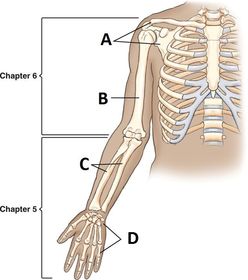
| A. Shoulder Girdle B. Humerus C. Radius & Ulna D. Hand & Wrist | |
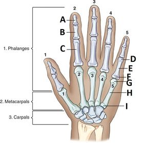
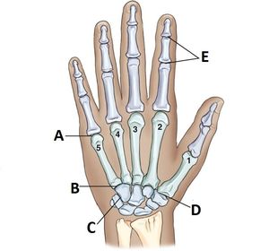
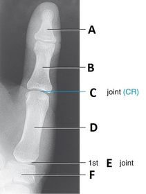
| A. Distal B. Middle C. Proximal D. Head E. Body F. Base G. Head H. Body I. Base | |
| A. Interphalangeal Joint (IP) B. 1st Metacarpophalangeal Joint (MCP) C. Trapezoid D. Trapezium E. Capitate F. Hamate G. 5th Carpometacarpal Joint (CMC) H. 5th Metacarpophalangeal Joint (MCP) I. Proximal Interphalangeal Joint (PIP) J. Distal Interphalangeal Joint (DIP) | |
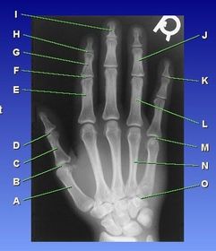
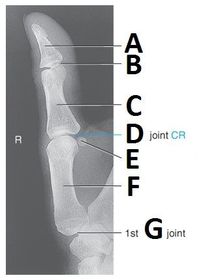
| Joint types and bone types | A. first metacarpal B. MP joint 1st digit C. proximal phalanx 1st digit D. IP joint 1st digit E. proximal phalanx 2nd digit F. PIP 2nd digit G. middle phalanx 2nd digit H. DIP 2nd digit I. distal phalanx 3rd digit J. middle phalanx 4th digit K. DIP 5th digit L. proximal phalanx 4th digit M. 5th MP joint N. 4th metacarpal O. 5th CM joint |
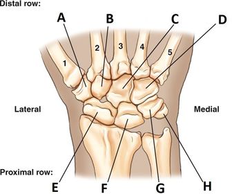
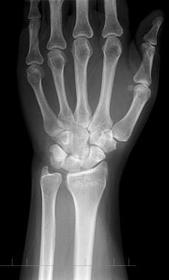
| A. Trapezium B. Trapezoid C. Capitate D. Hamate E. Scaphoid F. Lunate G. Triquetrum H. Pisiform | |
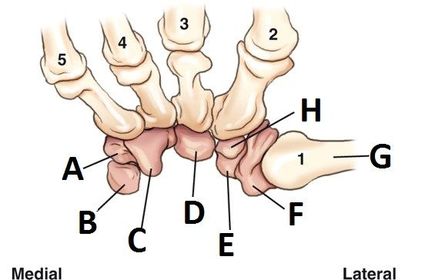
| A. Triquetrum B. Pisiform C. Hamate D. Capitate E. Scaphoid F. Trapezium G. thumb H. Trapezoid | |
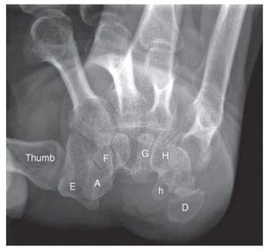
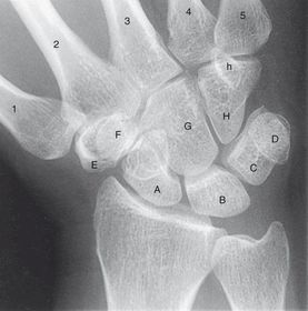
| A. Scaphoid D. Pisiform E. Trapezium F. Trapezoid G. Capitate H. Hamate h. Hamulus (process of Hamate) | |
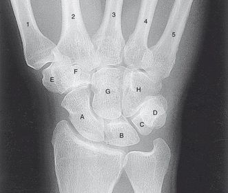
| A. scaphoid B. lunate C. triquetrum D. pisiform E. trapezium F. trapezoid G. capitate H. hamate | |
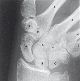
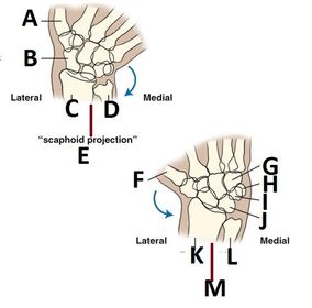
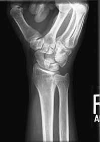
| Radial Deviation | A. scaphoid B. lunate C. triquetrum D. pisiform E. trapezium F. trapezoid G. capitate H. hamate |
| Ulnar Deviation | A. scaphoid B. lunate C. triquetrum D. pisiform E. trapezium F. trapezoid G. capitate H. hamate |
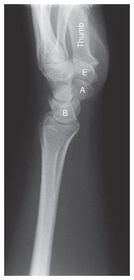
| Lateral Wrist | A. scaphoid B. lunate C. trapezium |
| Types of Joints | A. MCP - Metacarpophalangeal B. CMC - Carpometacarpal C. IC - Intercarpals D. CMC Carpometacarpal E. IP - Interphalangeal (proximal = PIP, distal = DIP) |
| A. thumb B. scaphoid C. radius D. ulna E. Ulnar Deviation F. thumb G. hamate H. pisiform I. triquetrum J. lunate K. radius L. ulna M. Radial Deviation | |
| What would displacement of this indicate? | Wrist Fat Pad displacement could indicate a fracture or more serious injury |
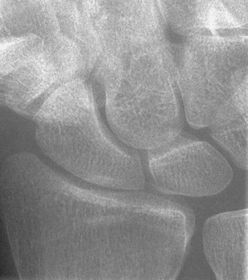
| What is the most commonly fractured carpal bone? | Scaphoid |
| Inflammation of the bursae, or fluid filled sacs, that enclose the joints is called _______ | Bursitis |
| The transfer of disease or cancerous lesions from one organ or part that may not be directly connected is called ____ ______ . Malignant tumors have the ability to metastasize, or transfer from one body part to another. | Bone Metastases |
| the painful disorder of the wrist and hand that results from compression of the median nerve is called ____ ____ syndrome. | Carpal Tunnel |
| Fracture and dislocation of the posterior lip of the distal radius is called ______ ______ . | Barton's Fracture |
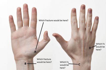
| A fracture of the base of the first metacarpal bone is called ____ ____ . | Bennett's Fracture |
| A transverse fracture that extends through the metacarpal neck - most commonly seen in the 5th metacarpal is called ________ . | Boxer's Fracture |
| A transverse fracture of the distal radius where the distal fragment is displaced posteriorly is called _____ _____ . | Colle's Fracture |
| the reverse of Colle's Fracture, the transverse fracture of the distal radius with the distal fragment displaced anteriorly is called __________ . | Smith's Fracture |
| A ____ _____ describes the accumulated fluid (synovial or hemorrhagic) in the joint cavity. | Joint Effusion |
| _____ AKA degenerative joint disease, is a noninflammatory joint disease where the bone gradually deteriorates | Osteoarthritis |
| A local or generalized infection of bone or bone marrow is called _______ . | Osteomyelitis |
| A hereditary disease marked by abnormally dense bone is called _____ | Osteopetrosis |
| The reduction in the quantity of bone, or atrophy of skeletal tissue is called _______ . | Osteoporosis |
| A common chronic skeletal disease where the bone is destructed and repaired by the overproduction of very dense yet soft bones that fracture easily is termed ______ . | Paget's Disease |
| A chronic systemic disease with inflammatory changes throughout the connective tissues is called ______ arthritis. It's more common in women than in men. | Rheumatoid Arthritis |
| ____ ____ refers to a sprain or tear of the ulnar collateral ligament of the thumb near the MCP joint | Skier's Thumb |
| Bone tumors or neoplasms are also called ____ ____ . | Bone Neoplasia |
| Neoplasms are also called ____ | Tumors |
| The most primary malignant cancerous bone tumor that affects people 40 - 70 years old is called ______ ______ . | Multiple Myeloma |
| The 2nd most common type of malignant primary cancerous bone tumor that generally affects people 10-20 years old is called ____ ____ . | Osteogenic Sarcoma |
| A common malignant bone tumor in children and adults that arises from bone marrow is called ____ ____ . | Ewing's Sarcoma |
| A. Bennet's Fx B. Smith's Fx C. Colle's Fx D. Boxer's Fx | |
| What are the routine exams for a finger AND thumb? | PA PA Oblique Lateral |
| What are the routine exams for the hand? | PA PA Oblique Lateral Fan or Lateral |
| What are the routine exams for the wrist? | PA PA Oblique Lateral |
| PA Fingers Technique & Evaluation Criteria | SID 40 in Analog 50-55 kV Digital 55-60 kV IR 8x10 non grid CR @ PIP joint must see distal, middle, and proximal phalanges; distal metacarpal, and joints |
| PA Oblique Finger Medial or Lateral Rotation | SID 40 in Analog 50-55 kV Digital 55-60 kV IR 8x10 in non grid CR @ PIP joint must see distal, middle, and proximal phalanges, distal metacarpal, and joints |
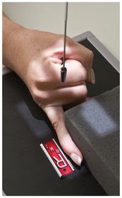
| Lateral Finger lateromedial or mediolateral | SID 40 in Analog 50 - 55 kV Digital 55 - 60 kV IR 8x10 in non grid CR @ PIP joint must see lateral view of distal, middle, and proximal phalanges; distal metacarpal, and joints. |
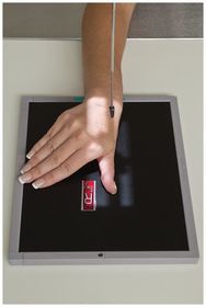
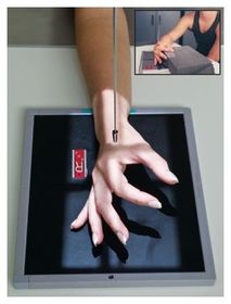
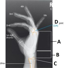
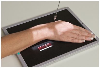
| AP Thumb | SID 40 in Analog 50 - 55 kV Digital 55 - 60 kV IR 8x10 in non grid CR @ 1st MCP joint must see distal and proximal phalanges, first metacarpal, trapezium, and associated joints IP and MC joints should appear open |
| PA Oblique Thumb Medial Rotation | SID 40 in Analog 50 - 55 kV Digital 55 - 60 kV IR 8x10 in non grid CR @ 1st MCP joint must see distal and proximal phalanges, first metacarpal, trapezium, and associated joints |
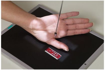
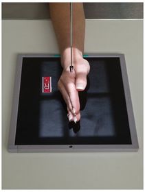
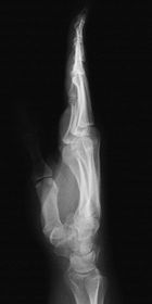
| Lateral Thumb | SID 40 in Analog 50 - 55 kV Digital 55 - 60 kV IR 8x10 in non grid CR @ 1st MCP joint must see distal and proximal phalanges, first metacarpal, trapezium (superimposed), and joints |
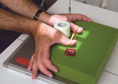
| AP Axial Thumb Modified Robert's Method | SID 40 in Analog 50 - 55 kV Digital 55 - 60 kV IR 8x10 in non grid CR angled 15 degrees proximally (toward wrist) @ 1st CMC joint |
| PA Stress | SID 40 in Analog 50 - 55 kV Digital 55 - 60 kV IR 8x10 in non grid CR midway between MCP joints |
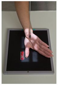
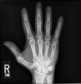
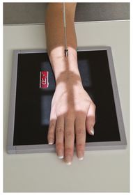
| PA Hand | SID 40 in Analog 50 - 55 kV Digital 55 - 60 kV IR 8x10 in nongrid CR @ 3rd MCP joint |
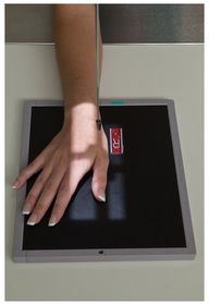
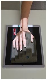
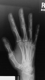
| PA Oblique Hand | SID 40 in Analog 50 - 55 kV Digital 55 - 60 kV IR 8x10 in nongrid CR @ 3rd MCP joint |
| Lateral Fan Hand Lateromedial | SID 40 in Analog 60-65 kV Digital 65-70 kV IR 8x10 in nongrid CR @ 2nd MCP joint |
| Lateral Hand Extension & Flexion | SID 40 in Analog 60-65 kV Digital 65-70 kV IR 8x10 in non grid CR @ 2nd to 5th MCP joints * alternative to fan lateral * |
| PA and AP Wrist | SID 40 in Analog 50 - 55 kV Digital 55 - 60 kV IR 8x10 in nongrid CR @ midcarpal area |
| PA Oblique Wrist | SID 40 in Analog 60-65 kV Digital 65-70 kV IR 8x10 in nongrid CR @ midcarpal area |
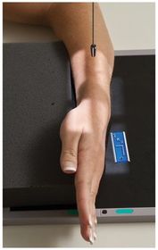
| Lateral Wrist Lateromedial | SID 40 in Analog 60-65 kV Digital 65-70 kV IR 8x10 in nongrid CR @ midcarpal area |
| PA Axial Ulnar Deviation Scaphoid Wrist | SID 40 in Analog 60-65 kV Digital 65-70 kV IR 8x10 in nongrid CR angled 10 - 15 degrees proximally centered at the scaphoid |
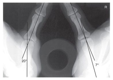
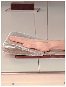
| PA Scaphoid-Hand Elevated Ulnar Deviation wrist | SID 40 in Analog 60 - 65 kV Digital 65 - 70 kV IR 8x10 in nongrid position and wrist palm down on IR with hand elevated on 20 degree sponge CR @ scaphoid |
| PA Radial Deviation Wrist | SID 40 in Analog 60 - 65 kV Digital 65 -70 kV IR 8x10 in nongrid CR @ midcarpal area |
| How many bones make up the hand and wrist? | 27 |
| Which carpal bone is the smallest? | Pisiform |
| the _______ position/projection is preferred for localizing foreign bodies | True Lateral |
| If the fingers are rotated how will the finger x-ray appear? | no spaces between the joints |
| for a PA Oblique finger, the fingers MUST be ______ to the IR | parallel |
| if you're doing an oblique x-ray of the 1st-3rd digit you're going to rotate the wrist _____ to reduce OID | medially |
| If you're doing an oblique x-ray of the the 4th-5th digit, you rotate the wrist _____ to reduce OID | laterally |
| For all of the fingers BESIDES the thumb, you are centering at the _____ joint. | PIP |
| For thumb projections you're going to center at the ______ joint. | MCP |
| the PA stress thumb x-ray demonstrates | tears in the ligaments |
| If the joints do not appear open on the x-ray, that means ______ | the hand was rotated, or fingers bent |
| For PA oblique of the hand, if the 4th and 5th digits are superimposed that means | the hand was not in a true 45 degree oblique |
| How can you tell if oblique position is good based on the radiograph of the 45 degree oblique finger? | the joints will be open and slightly concaved |
| It's better to see the _____ on the fan lateral position. | phalanges |
| Lateral hand, both fanned and true lateral, demonstrate the ______ fracture | Boxer's Fracture |
| true lateral hand position best shows ______ _____ in the hand | Foreign Bodies |
| AP Oblique Bilateral (ball catcher's) you should center at the ____ MCP joint, this radiograph demonstrates ______ | 5th rheumatoid arthritis, 5th metacarpal Fx's (commonly from punching) |
| For Boxer's Fx's AND 5th metacarpal Fx's, you need to ask the patient...... | what the cause of injury was |
| Why do you flex the fingers for the PA wrist? | to flatten the wrist for the projection |
| TTCH SLTP | Some Lovers Try Positions That They Can't Handle Scaphoid Lunate Triquetrium Pisiform Trapezium Trapezoid Capitate Hamate |
| For a lateral wrist you want to rotate the hand externally (laterally) __ degrees to get it in TRUE lateral | 5 degrees |
| For radial deviation projections you want to angle the CR ___ to ___ degrees | 10-15 degrees |
| For modified Stecher (modified ulnar deviation), you're elevating the hand ___ degrees on a sponge with no deviation. This is done only if patient cannot deviate the wrist. | 20 degrees |
| Which position should you use if the patient cannot angle their wrist for an Ulnar Deviation projection? | Modified Stecher |
| For most hand projections you should center the CR at the ___ ___ joint. Most lateral hand projections should be centered at the ___ ___ joint. | 3rd MCP joint 2nd MCP joint |
| TRUE OR FALSE? You want the forearm and elbow in the same plane as the finger, hand, or wrist that's being x-rayed. | TRUE |
| you should place your marker on the ____ side of the appendage in most cases. | LATERAL |
| TRUE OR FALSE? If you have to do several different appendage series all together, you should group them together. For example, all the AP's should be done first, then the PA's, then the obliques, etc... | TRUE |
| Why do you want to group all your similar projections together for multiple appendage projections? | Patient comfort Time efficiency |
| Bone Age Studies | Viewing growth plates in bones to observe the maturity and growth patterns |
| Which carpal bone is most anterior? | Pisiform |
| with the AP thumb projection, the hand is positioned in _____ ____ rotation. | Extreme Internal |
| Why is PA preferred vs AP in most hand, finger, and wrist projections? | PA reduces OID |
| Where do you place the CR for all finger exams? | PIP joint |
| Where do you place the CR for all thumb exams? | 1st MCP joint |
| Where do you place the CR for all hand exams? | 3rd MCP joint |
| Where do you put the CR for all wrist projections? | Midcarpals |
| What's the kVp range for finger projections? | 50-60 kVp |
| What's the kVp range for thumb projections? | 50-60 kVp |
| The kVp range for most hand projections is ___ - ___ kVp. The two projections that are still 50-60 kVp are ____ and ____ | 60-70 kVp PA Oblique and PA |
| What's the kVp range for wrist projections? | 60 - 70 kVp |
| For large plaster casts you ____ technique by __-___ kV. | increase 8-10 |
| For fiberglass casts you _____ technique by __-__ kV. | Increase 3-4 |
| _____ is a procedure that uses contrast injected into a joint capsule to visualize soft tissue pathology | .Arthrogram |
| You want to use a ____ focal spot for finger, hand and wrist projections | SMALL |
| You want ____ exposure time for finger, hand and wrist projections | SHORT |
| For the ulnar deviation projection, the CR should be angled ___ - ___ degrees ______ . | 10-15 proximally |
| Which projection/position demonstrates Rheumatoid Arthritis? | Norgaard "ball-catchers" AP Oblique Bilateral |
| Which projection/position demonstrates Carpal Tunnel Syndrome? | Carpal Canal Gaynor-Hart |
| Which projection/position demonstrates injuries to the Ulnar Collateral Ligament? | PA Stress Folio-method |
| Why is the AP projection preferred for the thumb instead of the PA? | Less OID |
| Modified Robert's | AP Axial Thumb |
| Stecher Method or Navicular View | Ulnar Deviation |
| Modified Stecher | Elevated Ulnar Deviation |
| Norgaard or Ball-Catcher's Method | AP Oblique Bilateral |
| Gaynor-Hart Method | Carpal Canal |
| Folio-Method | PA Stress Thumbs |
Want to create your own Flashcards for free with GoConqr? Learn more.