
Mind map
General Pathology
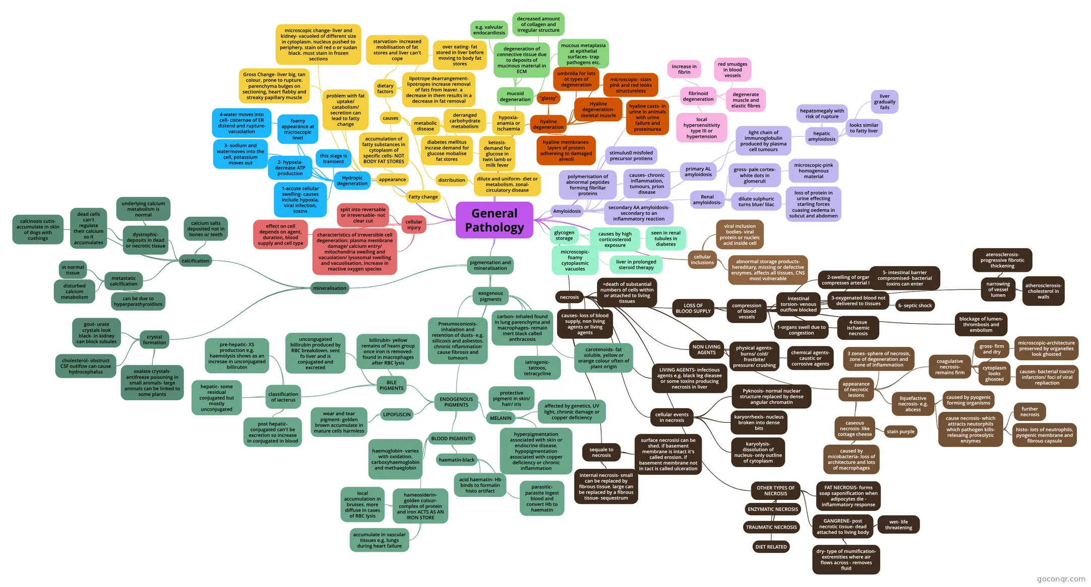
Mind map: General pathology
Map content
General Pathology
cellular injury
- split into reversable or irreversable- not clear cut
- effect on cell depends on agent, duration, blood supply and cell type
- characteristics of irreversible cell degeneration: plasma membrane damage/ calcium entry/ mitochondria swelling and vacuolation/ lysosomal swelling and vacuolisation. increase in reactive oxygen species
Hydropic degeneration
- 1-accute cellular swelling- causes include hypoxia, viral infection, toxins
- 2- hypoxia- decrease ATP production
- 3- sodium and watermoves into the cell, potassium moves out
- 4-water moves into cell- cisternae of ER distend and rupture- vacuolation
- foamy appearance at microscopic level
- this stage is transient
Fatty change
- appearance
- accumulation of fatty substances in cytoplasm of specific cells- NOT BODY FAT STORES
- problem with fat uptake/ catabolism/ secretion can lead to fatty change
- Gross Change- liver big, tan colour, prone to rupture. parenchyma bulges on sectioning, heart flabby and streaky papillary muscle
- microscopic change- liver and kidney- vacuoled of different size in cytoplasm. nucleus pushed to periphery. stain oil red o or sudan black. must stain in frozen sections
- causes
- dietary factors
- starvation- increased mobilisation of fat stores and liver can't cope
- over eating- fat stored in liver before moving to body fat stores
- lipotrope dearrangement- lipotropes increase removal of fats from leaver. a decrease in them results in a decrease in fat removal
- metabolic disease
- and toxins
- derranged carbohydrate metabolism
- diabetes mellitus incrase demand for glucose mobalise fat stores
- ketosis- demand for glucose in twin lamb or milk fever
- hypoxia- anamia or ischaemia
- distribution
- dilute and uniform- diet or metabolism. zonal- circulatory disease
- dietary factors
- appearance
mucoid degeneration
- degeneration of connective tissue due to deposits of mucinous material in ECM
- e.g. valvular endocardiosis
- decreased amount of collagen and irregular structure
- mucous metaplasia at epithelial surfaces- trap pathogens etc.
hyaline degeneration
- "glassy"
- umbrella for lots ot types of degeneration
- microscopic- stain pink and red looks structureless
- Hyaline degeneration- skeletal muscle
- hyaline casts- in urine in animals with urine failure and proteinurea
- hyaline membranes- layers of protein adhereing to damaged alveoli
fibrinoid degeneration
- increase in fibrin
- red smudges in blood vessels
- degenerate muscle and elastic fibres
- local hypersensitivity type III or hypertension
Amyloidosis
- polymerisation of abnormal peptides forming fibrillar proteins
- stimulus0 misfoled precursor protiens
- causes- chronic inflammation, tumours, prion disease
- primary AL amyloidosis
- light chain of immunoglobulin produced by plasma cell tumours
- secondary AA amyloidosis- secondary to an inflammatory reaction
- Renal amyloidosis-
- gross- pale cortex- white dots in glomeruli
- stain brown or yellow with iodine
- dilute sulphuric turns blue/ lilac
- microscopic-pink homogenous material
- loss of protein in urine effecting starling forces cuasing oedema in subcut and abdomen
- gross- pale cortex- white dots in glomeruli
- hepatic amyloidosis
- hepatomegaly with risk of rupture
- looks similar to fatty liver
- liver gradually fails
glycogen storage
- causes by high corticosteroid exposure
- microscopic- foamy cytoplasmic vacuoles
- seen in renal tubules in diabetes
- liver in prolonged steroid therapy
cellular inclusions
- viral inclusion bodies- viral protein or nucleic acid inside cell
- location indicates virus types
- abnormal storage products- hereditary, missing or defective enzymes, affects all tissues, CNS most vulnerable
- viral inclusion bodies- viral protein or nucleic acid inside cell
necrosis
- =death of substantial numbers of cells within or attached to living tissues
- causes- loss of blood supply, non living agents or living agents
- LOSS OF BLOOD SUPPLY
- compression of blood vessels
- intestinal torsion- venous outflow blocked
- 5- intestinal barrier compromised- bacterial toxins can enter
- 1-organs swell due to congestion
- 2-swelling of organs compresses arterial flow
- 3-oxygenated blood not delivered to tissues
- 4-tissue ischaemic necrosis
- 6- septic shock
- intestinal torsion- venous outflow blocked
- narrowing of vessel lumen
- aterosclerosis- progressive fibrotic thickening
- atherosclerosis- cholesterol in walls
- blockage of lumen- thrombosis and embolism
- e.g. hypertrophic cardiomyopathy
- compression of blood vessels
- NON LIVING AGENTS
- physical agents- burns/ cold/ frostbite/ pressure/ crushing
- chemical agents- caustic or corrosive agents
- LIVING AGENTS- infectious agents e.g. black leg dieasee or some toxins producing necrosis in liver
- appearance of necrotic lesions
- 3 zones- sphere of necrosis, zone of degeneration and zone of inflammation
- coagulative necrosis- remains firm
- gross- firm and dry
- microscopic-architecture preserved by organelles look ghosted
- cytoplasm looks ghosted
- causes- bacterial toxins/ infarction/ foci of viral repliaction
- liquefactive necrosis- e.g. abcess
- caused by pyogenic forming organisms
- cause necrosis- which attracts neutorphils which pathogen kills- relesaing proteolytic enzymes
- further necrosis
- histo- lots of neutrophils, pyogenic membrane and fibrous capsule
- caseous necrosis- like cottage cheese
- like halfway between two
- caused by micobacteria- loss of architecture and lots of macrophages
- stain purple
- cellular events in necrosis
- Pyknosis- normal nuclear structure replaced by dense angular chromatin
- karyorrhexis- nucleus broken into dense bits
- karyolysis- dissolution of nucleus- only outline of cytoplasm
- sequale to necrosis
- surface necrosisi can be shed. if basement membrane is intact it's called erosion. if basement membrane not in tact is called ulceration
- internal necrosis- small can be replaced by fibrous tissue. large can be replaced by a fibrous tissue- sequestrum
- OTHER TYPES OF NECROSIS
- FAT NECROSIS- forms soap saponification when adipocytes die - inflammatory response
- ENZYMATIC NECROSIS
- TRAUMATIC NECROSIS
- DIET RELATED
- GANGRENE- post necrotic tissue- dead attached to living body
- wet- life threatening
- dry- type of mumification- extremities where air flows across - removes fluid
pigmentation and mineralisation
- exogenous pigments
- inhaled, ingested and iatrogenic
- carbon- inhaled found in lung parenchyma and macrophages- remain inert black called anthracosis
- Pneumoconiosis- inhalation and retention of dusts- e.g. sillicosis and asbestos. chronic nflammation cause fibrosis and tumours
- carotenoids- fat soluble, yellow or orange colour often of plant origin
- iatrogenic- tattoos, tetracycline
- ENDOGENOUS PIGMENTS
- MELANIN
- protective pigment in skin/ hair/ iris
- affected by genetics, UV light, chronic damage or copper deficiency
- hyperpigmentation associated with skin or endocrine disease. hypopigmentation associated with copper deficiency or chronic inflammation
- BLOOD PIGMENTS
- haemoglobin- varies with oxidation. carboxyhaemoglobin and methaeglobin
- hameosiderin- golden colour- complex of protein and iron ACTS AS AN IRON STORE
- local accumulation in bruises. more diffuse in cases of RBC lysis
- accumulate in vascular tissues e.g. lungs during heart failure
- haematin-black
- acid haematin- Hb binds to formalin histo artifact
- parasitic- parasite ingest blood and convert Hb to haematin
- LIPOFUSCIN
- wear and tear pigment- golden brown accumulate in mature cells harmless
- BILE PIGMENTS
- billirubin- yellow remains of heam group once iron is removed- found in macrophages after RBC lysis
- uncongugated billirubin produced by RBC breakdown. sent fo liver and is conjugated and excreted
- classification of iacterus
- pre-hepatic- XS production e.g. haemolysis shows as an incresae in unconjugated billirubin
- hepatic- some residual conjugated but mostly unconjugated
- post hepatic- conjugated can't be excretion so increase in conjugated in blood
- MELANIN
- mineralisation
- calcification
- calcium salts deposited not in bones or teeth
- dystrophic- deposits in dead or necrotic tissue
- underlying calcium metabolism is normal
- dead cells can't regulate their calcium so it accumulates
- metastatic calcification
- in normal tissue
- disturbed calcium metabolism
- can be due to hyperparathyroidism
- calcinosis cutis- accumulate in skin of dogs with cushings
- crystal formation
- gout- urate crystals look black- in kidney can block tubules
- cholesterol- obstruct CSF outlfow can cause hydrocephalus
- oxalate crystals- antifreeze poisoning in small animals- large animals can be linked to some plants
- calcification
- exogenous pigments
Information
- Description
- Degree level Year 2 (principles of science) Mind Map on General Pathology, created by Clodagh Mullins on 13/12/2017.
- Created by
- Clodagh Mullins
- Views
- 5
- Rating