297732
Description
Mind Map by melian.yates, updated more than 1 year ago
|
|
Created by melian.yates
over 10 years ago
|
|
Granulomatous Disease
- Classifications
- With/ Without Granulomas
- All Granulomas are part
of Granulomatous
inflammation
- Not all granulomatous
inflammatory lesions
have granulomas
- Granuloma = Focal/ multifocal
- Granulomatous = Thickened/ Turgid
- Granulomatous = Thickened/ Turgid
- Not all granulomatous
inflammatory lesions
have granulomas
- All Granulomas are part
of Granulomatous
inflammation
- Cells Present
- 1) Granulomatous
- Macrophages
- Multinucleated giant
cells (Macrophages),
Lymphocytes,
Fibroblasts
- Multinucleated giant
cells (Macrophages),
Lymphocytes,
Fibroblasts
- Macrophages
- 2) Eosinophilic Granulomatous
- Macrophages + Eosinophils
- Cats & horses
- Ex. Feline
Eosinophilic
granuloma complex
- Ex. Feline
Eosinophilic
granuloma complex
- Macrophages + Eosinophils
- 3) Pyogranulomatous
- Macrophages + Neutrophils
- Ex. Actinobacillus ligieresii,
Actinomyces bovis
- Ex. Actinobacillus ligieresii,
Actinomyces bovis
- Macrophages + Neutrophils
- 1) Granulomatous
- Aetiology
- Bacteria
- Actinobacillus lignieresii
- Actinomyces bovis
- Actinomyces bovis
- Live in Macrophages:
- Mycobacteria
- M. tuberculosis complex
- M. boivs
- Cattle,
other
animals
- Spread by
coughing &
sneezing
- Survive in
respiratory
tract macrophages
- Survive in
respiratory
tract macrophages
- < 10% of infected
animals develop
clinical disease
- Zoonotic
- Pathology
- Gross
- Well circumscribed,
often encapsulated,
pale yellow to white
foci, often with
Caseous Necrosis
&/or mineralization
- Most often
respiratory tract
lymph nodes are
affected
- Can have
lesion in lungs
- Can have
systemic
disease
- Granulomas all over body
- Granulomas all over body
- Well circumscribed,
often encapsulated,
pale yellow to white
foci, often with
Caseous Necrosis
&/or mineralization
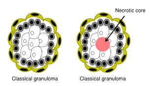
- Histologically
- Central area of
Caseous Necrosis
- Classical Granulomas
- Acid fast bacteria w/in
internal macrophages &
giant cells
- Acid fast bacteria w/in
internal macrophages &
giant cells
- Central area of
Caseous Necrosis
- Gross
- Cattle,
other
animals
- M. boivs
- (Cats) M. lepraemurium
- Cutaneous nodular lesions
- Refractory to normal Abx
- Granulomatous
dermatitis with Acid
fast organisms
- Refractory to normal Abx
- Cutaneous nodular lesions
- M. avium complex (MAC)
- M. a paratuberculosis
- Johne's disease
- Cattle, sheep, goats, deer
- Animals infected when young
- Contaminated feces
- Bacteria lives in
macrophages for years: subclinical
- Causes diarrhaoea &
weight loss when older
- Causes diarrhaoea &
weight loss when older
- Contaminated feces
- Animals infected when young
- Pathology
- Grossly
- Thickened &
corrugated
small intestine
- Can be yellow in Sheep
- Terminal ileum is
most often affected
- Iliocecal junction
- Iliocecal junction
- Can be yellow in Sheep
- Enlarged
mesenteric
lymph nodes
- Thickened &
corrugated
small intestine
- Histologically (Sheep)
- Multibacillary
- Macrophages
& Giant cells
- Many Intracellular
acid fast bacteria
- Macrophages
& Giant cells
- Paucibacillary
- Lymphocytes &
Plasma cells w/
Macrophages
- Very few
acid fast
bacteria
- Rare in Cattle
- Lymphocytes &
Plasma cells w/
Macrophages
- Both types have
the same clinical
signs, gross lesions
& outcome
- Multibacillary
- Grossly
- Cattle, sheep, goats, deer
- Johne's disease
- M.a. avium
- Avian mycobacteriosis
- Granulomas in liver,
intestines & other
organs
- Granulomas in liver,
intestines & other
organs
- M. a paratuberculosis
- Pathogenesis
- Intracellular survival
aided by specialized
cell wall
- Stop the
lysosome
from fusing
- Capsular
polysaccharide,
complex free lipids
- Mycolic Acids
- Arabinogalactan
- Peptidoglycan
- Peptidoglycan
- Arabinogalactan
- Mycolic Acids
- Stop the
lysosome
from fusing
- Intracellular survival
aided by specialized
cell wall
- M. tuberculosis complex
- Rhodoccocus
- Brucella
- Salmonella
- Salmonella
- Brucella
- Stop phagolysosome fusing
- Inhibit lysosome enzymes
- Capsule resistant
to lysosomal
enzymes
- Capsule resistant
to lysosomal
enzymes
- Inhibit lysosome enzymes
- Mycobacteria
- Actinobacillus lignieresii
- Parasites
- Lungworm
- Cat: Aelurostrongylus abstrusus
- Sheep: Muellerius capillaris
- Cattle: Dictyocaulus
Viviparus
- Dog: Angiostrongylus vasorum
- Cat: Aelurostrongylus abstrusus
- Lungworm
- Fungi
- Aspergillus sp.
- Opportunistic
- Immunocompromised
animals
- Viral infection of gut
- Antibiotic therapy
- Antibiotic therapy
- Viral infection of gut
- Immunocompromised
animals
- Granulomas &
Pyogranulomas
- Opportunistic
- Systemic Mycoses
- Coccidioides immitis
- Inhalation
- Granulomas &
Pyogranulomatous
- Granulomas &
Pyogranulomatous
- Inhalation
- Coccidioides immitis
- Aspergillus sp.
- Foreign Material
- Most foreign material
will induce a
granulomatous
response
- Difficult/ Impossible for
Macrophages to
breakdown foreign
material
- Suture Material
- Silica
- Hair shafts ( Furunculosis)
- Sperm granuloma
- Sperm granuloma
- Hair shafts ( Furunculosis)
- Silica
- Difficult/ Impossible for
Macrophages to
breakdown foreign
material
- Most foreign material
will induce a
granulomatous
response
- Viruses
- Only 2 Viruses
- FIPV (Feline
Infectious Peritonitis
Virus)
- Coronavirus
- Feline infectious peritonitis (FIP)
- Lives in macrophages
- Vasculitis
- Wet & Dry forms of disease
- Wet
- Abdominal distension
- Dyspnea due
to thoracic
effusion
- Dyspnea due
to thoracic
effusion
- Abdominal distension
- Dry
- Most often
involves the eyes
& CNS
- Areas of
inflammation on
Retina
- Areas of
inflammation on
Retina
- Kidney, Liver, lymph nodes
- Most often
involves the eyes
& CNS
- Wet
- Infected Macrophages
accumulate around
small blood vessels
- Deposition of immune
complexes & Activation of
Complement
- Macrophages release
proinflammatory
cytokines which promote
leakage of blood vessels
- Damage to Blood vessels
- Damage to Blood vessels
- Deposition of immune
complexes & Activation of
Complement
- Wet & Dry forms of disease
- Vasculitis
- FECV => FIPV
- Clinical Signs
- Malaise, fluctating
fever, inappetance,
weight loss
- Malaise, fluctating
fever, inappetance,
weight loss
- Lives in macrophages
- Closely related
to FECV -
Harmless
- Feline infectious peritonitis (FIP)
- Coronavirus
- PCV-2 ( Porcine
circovirus 2)
- Circovirus
- PCVAD (PCV -2
Associated Disease)
- Systemic Disease
- PMWS (Post weaning
multisystemic wasting
syndrome)
- PMWS (Post weaning
multisystemic wasting
syndrome)
- Respiratory, Enteric,
Reproductive Disease
- Porcine Dermatitis
and Nephropathy
Syndrome (PDNS)
- Systemic Disease
- Not all infected pigs develop disease
- Granulomatous Inflammation
- Lymph nodes
- Lungs
- Intestines
- Liver
- Other tissues
- Other tissues
- Liver
- Intestines
- Granulomatous
lymphadenitis
- Lungs
- Lymph nodes
- Circovirus
- FIPV (Feline
Infectious Peritonitis
Virus)
- Only 2 Viruses
- Toxins
- Hairy Vetch (Vicia villosa) - Legume
- Causes necrosis &
granulomatous inflammation
in many organs (Heart, Kidney, Liver)
- Causes necrosis &
granulomatous inflammation
in many organs (Heart, Kidney, Liver)
- Hairy Vetch (Vicia villosa) - Legume
- Idiopathic
- GME (Granulomatous
meningoencephalomyelitis
of Dogs)
- Histiocytic ulcerative colitis
- Dogs: Sebaceous adenitis
- Feline eosinophilic
granuloma complex
- Consists of 3 diseases
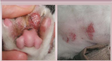
- Feline Eosinophilic Granuloma
- More common in young cats
- Nodular or linear lesions
on skin, footpad,
mucotaneous junctions &
oral cavity
- Raised, pink, alopecic
- Histology: Eosinophils &
Macrophages
- Raised, pink, alopecic
- Nodular or linear lesions
on skin, footpad,
mucotaneous junctions &
oral cavity
- More common in young cats
- Eosinophilic plaque
- Raised, red, alopecic
to ulcerated, flat topped
plaques on the skin
- VERY Pruritic
- Eosinophils,
Lymphocytes &
Macrophages
- Eosinophils,
Lymphocytes &
Macrophages
- VERY Pruritic
- Raised, red, alopecic
to ulcerated, flat topped
plaques on the skin
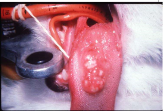
- Indolent Ulcer
- Ulcerated lesion on
upper lip adjacent to the
philtrum
- Pain & pruritis are rare
- Unilateral or Bilateral
- Histologically:
Eosinophils,
Neutrophils, Mast cells
& Macrophages
- Histologically:
Eosinophils,
Neutrophils, Mast cells
& Macrophages
- Unilateral or Bilateral
- Pain & pruritis are rare
- Ulcerated lesion on
upper lip adjacent to the
philtrum
- Feline Eosinophilic Granuloma
- Consists of 3 diseases
- Feline eosinophilic
granuloma complex
- Dogs: Sebaceous adenitis
- Young to middle
aged small dog
breeds
- Causes
neurological
disease
- Can affect spinal
cord, brain
meninges
- Can affect spinal
cord, brain
meninges
- Causes
neurological
disease
- Gross Lesions
- Few (maybe some
discoloration)
- Few (maybe some
discoloration)
- Histologically
- Inflammation &
Necrosis of the
white matter
- Macrophages,
Lymphocytes,
Plasma cells
- Inflammation &
Necrosis of the
white matter
- Histiocytic ulcerative colitis
- GME (Granulomatous
meningoencephalomyelitis
of Dogs)
- Bacteria
- With/ Without Granulomas
Media attachments
Want to create your own Mind Maps for free with GoConqr? Learn more.