5187499
Description
Mind Map by Lindie Metz, updated more than 1 year ago
|
|
Created by Lindie Metz
almost 10 years ago
|
|
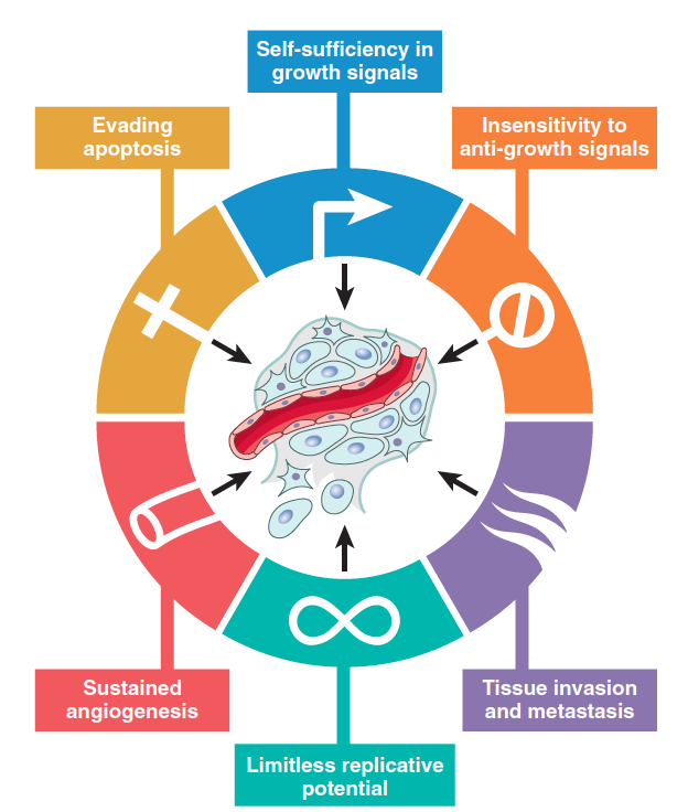
7 Fundamental
Changes
- Insensitivity to growth inhibitory
signals
- RB tumour suppressor
- RB enforces gap btw G0 and G1 – determines if cell can continue
- Recessive gene, so require homozygous allele mutation
- >> Retinoblastoma = interocular cancer (+ breast, bladder, lung)
- DNA viruses can render Rb non-functional (HPV, HBV, EBV)
- Binds to Rb and prevents functioning
- Binds to Rb and prevents functioning
- RB enforces gap btw G0 and G1 – determines if cell can continue
- p53 gene
- Guardian of the genome!
- Tumour supressor gene STOP neoplasm
- Activates quiescence (temp cell arrest)
- Activates senescence (permanent)
- Triggers apoptosis
- Activates quiescence (temp cell arrest)
- Monitors cell stress and directs best response
- Homozygous loss of p53
- DNA damage unrepaired
- Mutations remain in dividing cells
- Malignant transformation
- DNA damage unrepaired
- DNA viruses can render Rb non-functional (HPV, HBV, EBV)
- Guardian of the genome!
- RB tumour suppressor
- Tissue invasion and
apoptosis
- Invasion of ECM
- ECM = BM + interstitial CT
- 1. Detach tumour cells from oneathother
- 2. Degrade ECM and ICT
- 3. Change attachment of tumour cells to ECM proteins
- 4. Migration of tumour cells through degraded BM
and ICT directed by tumor cell derived cytokines
- See diagram!
- ECM = BM + interstitial CT
- Vascular dissemination and
homing of tumour cells
- Tumour cells in circulation are vulnerable to host immunity
- 1. Attach to leuk/platelets > emboli >
protected from anti tumour host cell
- 2. Extravasation – tumour cells/emboli attach to EN
>> through BM >> into parenchyma
- Predict site of extravasation/metastasis
- Location of primary tumour
- Vascular/lymph drainage of primary tumour
- Location of primary tumour
- Tumour cells in circulation are vulnerable to host immunity
- Invasion of ECM
- Self-sufficiency in growth signals
- Oncogenes promote autonomous Ca cell growth
- > Promoted out of quiescent stage with no add stimulus
- > By synthesizing GF + generate constant mitotic signals to cell
- > Autonomous and disregulated
- Oncogenes promote autonomous Ca cell growth
- Limitless replicative potential
- Normal cells lose capacity to divide after 60/70 times
- = sensence d/t shortened telomeres (p53/Rb)
- p53/Rb checkpoints disabled so Ca must overcome mitotic catastrophe
- Tumour cell reactivates telomerase >> activates stem cells
- Tumour cell reactivates telomerase >> activates stem cells
- Telomerase maintenance in 85-95% Ca
- >> Break cycle of fusion and breakage
- >> Break cycle of fusion and breakage
- >> Avoid cell death
- Normal cells lose capacity to divide after 60/70 times
- Sustained angiogenesis
- Tumour needs nutrients/O2 + waste removal
bigger than 1-2mm > products can diffuse
- = neo-angiogenesis + vasculogenesis >> new vessels
from capillaries + endothelial cells from bone marrow
- BUT vessels are abnormal and leaky
- Supports tumour growth
- O2/nutrient supply + waste removal
- EN cells stimulate new tumour cell growth by GF
- >> supplies new channels for metastases
- O2/nutrient supply + waste removal
- Tumour needs nutrients/O2 + waste removal
bigger than 1-2mm > products can diffuse
- Evading apoptosis
- Inc in neoplastic cells + mutations
in genes that regulate apoptosis
- >> Inc in cells + << in apoptosis
- Inc in neoplastic cells + mutations
in genes that regulate apoptosis
Media attachments
Want to create your own Mind Maps for free with GoConqr? Learn more.