
Notes
AS biology
Content of the notes (3)
Page 1
cells.
*light microscope-(M *1500) (R 200 NM) they have low resolution compared to electron microscope.
*Electron microscope- (R 0.1nm) magnification for (Transmission EM= *500,000 creates 2D image) (scanning EM= 100,000 creates 3D image)
*Magnification-the degree to which the size of an image is larger than the object itself.
*Resolution-the degree to which it is possible to distinguish between 2 objects that are very close together.
*Staining samples for light microscope:
Staining- coloured stains are chemicals which binds to the chemicals on/in the specimen, allowing them to be seen.
Sectioning-specimen are embedded in wax and thin sections are sliced without destroying cell structures.useful for sampling sections of soft tissue.
*electron microscope :
Staining- electron microscope starts off black and white, the colour is added afterwards using specialised computer program.
(TEM) transmission electron microscope - electron beam passes through thin sample, electron can't pass through the denser part of the sample creating contrast.Final image is 2D
(SEM) scanning electron microscope - electron beam directed onto the sample but doesn't pass through the specimen they "bounced off" the sample.final Imagen is 3D and the surface of the sample.
ADVANTAGE- The resolution is 0.1 nm which is more than the light microscope this can create more detailed image of the structure (organelles) in cell. (SEM) produces 3D image which revels detail of tissue arrangement, which isn't possible using light microscope.
LIMITATION - Electron beans deflected by molecules in the air, so samples have to be placed in a vacuum. electron microscopes are very expensive and preparing the sample requires high degree of skill and training.
* Cytoskeleton- Are a network of fibres made of protein. which keep the cells shape stable by providing an internal framework which is the cytoskeleton. The types of fibre:
microfilaments-small solid strands e.g. actin are present in the muscles cells can cause movement seen in white blood cells and they also move organelles around inside cells.
Microtubules- cylinders about 25 nm made of protein called tubulin, these fibres can be used to move organism through liquid or waft liquid past cell
OVERALL FUNCTION- Support organelles / strengthen the cell and maintain cell shape / transport materials within cells / cell movement. uses ATP.
* FLAGELLA (UNDULIPODIA) & CILIA.- They are structurally the same, they are like extensions which stick out of the cell surface. Each made up of a cylinder containing 9 microtubules arranged in a circle. The undulipodium which forms the tail of the sperm moves the whole cells, on the protoctista it enables it to move.
BIOLOGICAL MEMBRANES/ FLUID BOUNDARIES.
Membrane surrounds all cells are partially permeable barriers which separates the content of the cell form the outside world, the roles:
* Separates cell contents from the outside environment
*separates cell components form cytoplasm
*cell recognition and cell signaling
*regulating the transport of materials into and out of the cells.
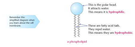
*Cell/ plasma membranes basic structural compound is the phospholipid bilayer (NOT SEMI- PERMEABLE BUT PARTIALLY PERMEABLE)
*The phosphate head is HYDROPHILIC.(WATER LOVING)
*Two fatty acid tail are HYDROPHOBIC.(WATER HATING)
* All membrane are permeable to water molecule as it can diffuse through the lipid bilayer (more permeable means more aquaporin which is the protein channel which lets water in)
THE FLUID MOSAIC MODEL
* This refers to the model of cell membrane structure, the lipid molecule gives it fluidity and the protein in the membrane gives it a mosaic appearance. The fluid mosaic contains:
CHOLESTEROL -gives membrane of the eukaryotic cell stability and fluidity. It fits between fatty acid tails to make the barrier more complete.
CHANNEL/ CARRIER PROTEIN - Is present of the membrane transport functions, the channel proteins allow movement of large (eg Glucose) or hydrophilic molecule to just pass through the bilayer. The carrier proteins actively move substances across the membrane.
RECEPTOR SITES- Used for recognition & communication. Allows some hormones to bind with cells so that a cell response can be carried out.
GLYCOPROTEINS & GLYCOLIPIDS- Also used for recognition & communication, this shows the cells are themselves. they can also bind cells together in tissues.
*MEMBRANES & TEMPERATURE- increasing the temperature gives molecules more kinetic energy so they move faster. this increases movement of phospholipids and other components this makes membranes leaky which allows substances that would normally not do so to enter or leave the cell.
Page 2
Organelles Structure and function:
UNDER ELECTRON MICROSCOPE.
ORGANELLES SURROUNDED BY MEMBRANES:
* THE NUCLEUS- Nucleus is the largest organelle which is seen.darker patch when stained is the chromatin. this is surrounded by nuclear envelope nuclear pores go through the envelope there is a large dense spherical structure called nucleous.
- the function is that it has all the cells genetic material, the chromatin consists of DNA and proteins. this has instructions for making proteins some of which regulate cells activity. Also makes ribosome and RNA.
* ENDOPLASMIC RETICULUM (ER)- series of flattened, membrane bound scales called cisternae, continuous with outer nuclear membrane (RER) surrounded by ribosomes (SER) doesn't have ribosomes.
-The functions :
The (RER) transports proteins made on the attached ribosomes.some of these proteins can be secreted from the cells and some placed on the cell surface membrane.
The (SER) is involved in making the lipids that the cell needs.
*GOLGI APPARATUS- A stack of membrane bound flattened sacs
- Functions they receive thee protein from the RER and modifies them, may add more sugar molecules. Then it packages the modified proteins into vesicles which can be transported.some modified cells move to the cell surface so it can be secreated out.
* MITOCHONDRIA (sing: mitochondrion)- can be spherical or sausage-shaped.they have 2 membranes separated by fluid-filled space. The inner membrane is folded to form cristae. central part of mitochondria is the matrix.
-Function, this is the site where (ATP) ADENOSINE TRIPHOSPHATE is produced during respiration. ATP is called a universal energy carrier as all activity which needs energy is provided by ATP.
*CHLOROPLAST- ONLY FOUND IN PLANT CELLS AND CELLS OF SOME PROTOCTISTS They are separated by fluid filled space. inner membrane is continuous with thylakoids. stacks of thylakoids are called granum (plural:grana).
- Function this is the sight of photosynthesis in plant cells, light energy is used to drive this reaction, in which CARBOHYDRATE molecules are made form CO2 and H2O.
*LYSOSOME- They are spherical sacs surrounded by a single membrane
-Functions they contain powerful digestive ENZYMES. There role is to breakdown materials (eg, white blood cells). They help breakdown invading microorganisms: specialised lysosome (ACROSOME) In the head of sperms help penetrate the egg by breaking down the materials surrounding the egg.
ORGANELLES WITHOUT MEMBRANES SURROUNDING THEM:
*RIBOSOMES- They are tiny organelles some in cytoplasm and some attached to RER, each consisting to 2 subunit.
-Functions they are site of protein synthesis in a cell, act as assembly line where mRNA (coded information) from nucleus used to assemble proteins from amino acids.
*CENTRIOLES- They are small tubes of protein fibers (microtubules), there is a pair of them next to the nucleus on animal cells and in cells of some protoctists.
-Functions they take part in cell division, they form fibers known as SPINDLE which moves the chromosomes during nuclear division.
Page 3
ORGANELLES AT WORK
* ORGANELLES IN PRODUCTION AND SECRETION OF PROTEINS- The DNA contains the instructions, this is copied in the nucleus to a mRNA, this leaves the nucleus through the nuclear pore and then attaches to the ribosome ( can be the one in the RER). The ribosome then reads the instruction and used the codes to s or protein. assembled protein in RER is transported to the GOLGI APPARATUS in a vesicle, here it's modified and packaged so it's ready for release, this is again packed in a vesicle and moved to the surface where it fuses with the membrane and is secreted out of the cell.
PROKARYOTES AND EUKARYOTES
*Eukaryotic cells have a nucleus- They contain organelles some are membrane bound, these give cells complicated internal structure
*prokaryotic cells don't have a nucleus- They are bacterias which can cause disease but are also used in the industry (eg making yoghurt)and are much smaller than Eukaryotic cells. They have:
one membrane
No membrane bound organelles such as mitochondria & chloroplast
contain cell walls made of peptidoglycan (NOT CELLULOSE)
DNA is in cytoplasm ( circular DNA/ Bacterial chromosome)
DNA not surrounded by membrane ( nuclear envelope)
ATP produced in a specialised infolded region called MESOSOME.
Some prokaryotic cell have FLAGELLA (Function like undulipodia but have different internal structure)
Information
- Description
- Note on AS biology, created by sajanarai on 01/05/2016.
- Created by
- sajanarai
- Views
- 1
- Rating