
Mapa mental
Endocrine Pathology
Conteúdo do mapa
Endocrine Pathology
Pathogenic Mechanisms of Endocrine Disease
- 1) Primary Gland Hypofunction
- Destruction of secretory cells by a disease process (e.g. immune mediated or other inflammatory injury in thyroids, parathyroids or adrenal cortex)
- Failure of Gland development (Hypoplasia or agenesis)
- Biochemical defect in synthetic pathway of the hormone (usually genetic aberrations & not commonly identifed in vet species)
- 2) Secondary Gland Hypofunction
- Destruction of glandular tissue or pituitary leading to failure of secretion of trophic hormone
- => Hypofunction of target organ
- Ex. Endochronologically inactive pituitary neoplasms interefere w/ secretion & release of ACTH, TSH & FSH
- => Clinically detectable hypofunction of adrenal cortex, thyroid follicular cells & gonads => Ensuing atrophy
- Ex. Endochronologically inactive pituitary neoplasms interefere w/ secretion & release of ACTH, TSH & FSH
- => Hypofunction of target organ
- Destruction of glandular tissue or pituitary leading to failure of secretion of trophic hormone
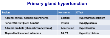
- 3) Primary Gland Hyperfunction
- One of the Most Common
- Hyperplasia/neoplasia of endocrine cells is involved
- The primary source synthesises & secretes hormone autonomously at a rate in excess of the ability of the body to utilise & degrade th ehormone
- 4) Secondary Gland Hyperfunction
- Lesion in one gland causes excessive trophic hormone secretion which results in long-term hypersecretion in a target organ
- Ex. Canine ACTH-secreting pituitary tumor => Adrenal cortical hyperplasia/hypertrophy & Hypercorticolism (Canine Cushing's Syndrome)
- Lesion in one gland causes excessive trophic hormone secretion which results in long-term hypersecretion in a target organ
- 5) Hypersecretion of Hormones or Hormone-like factors by non-endocrine tumors
- Ex. Adenocarcinoma of the anal sac apocrine glands in Dogs
- Produces parathyroid hormone-related peptide (PTHRP) => pseudo-hyperparathyroidism => Hypercalcaemia
- Ex. Some canine lymphomas (lymphosarcomas) may also exhibit a similar effect (i.e. the tumor secretes or is associated w/ a factor which induces hypercalcaemia)
- Ex. Adenocarcinoma of the anal sac apocrine glands in Dogs
- 6) Failure of Target cell response
- May be a failure of the cell surface receptor mechanism
- Lack of a 2nd intracellular messenger (e.g. cAMP) response
- Ex. Diabetes mellitus (Insulin resistance in Obesity - receptor loss in connective tissue cells, which then fail to react to insulin)
- 7) Endocrine Hyperactivity 2ndary to disease in other organs
- Disease in one organ results in hyperfunction of an endocrine gland
- Ex. 2ndary hyperparathyroidism associated w/ chronic renal failure
- Retention of phosphorous & alterations in Vit. D metabolism lead to hypercalcaemia, w/ resultant hyperparathyroidism & skeletal demineralization
- 8) Abnormal Hormone Degradation
- Decreased degradation: Can manifest as Hypersecretion
- Increased degradation: May relate to chronic drug administration
- 9) Iatrogenic Causes
- Long term administration of hormones may lead to direct or indirect effects
- Ex. Corticosteroid excess -> Cortisol -> muscle weakness, calcinosis cutis, hair loss & adrenal cortical atrophy
- 1) Primary Gland Hypofunction
Pituitary Gland
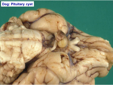
- Pituitary Cysts
- Pituitary Dwarfism (Juvenile panhypopituitarism)
- Most common in German Shepards
- Most cases: Due to failure of the oropharnygeal ectoderm to differentiate into pars distalis
- Associated w/ development of a pituitary cyst
- Signs become apparent at 2 months of age: Retention of puppy coat progressing to bilaterally symmetrical alopecia & hyperpigmented skin
- Associated w/ development of a pituitary cyst
- Pituitary Dwarfism (Juvenile panhypopituitarism)
- Neoplasia (Pituitary)
- ACTH secreting adenomas
- Most common in the Dog
- Leads to syndrome of cortisol excess (Cushing's syndrome)
- Pars intermedia neoplasia (Horse)
- Although some tumors are endochrinologically active, many of the features in equine disease are associated w/ the compression effect on the hypothalamus by the pituitary neoplasm
- Affects: seasonal shedding of hair, appetite, body temperature
- Non-functional pituitary neoplasms
- Combination of atrophy of surrounding pituitary & local extension into the brain can cause a range of problemms
- => Weakness, collapse, blindness
- Combination of atrophy of surrounding pituitary & local extension into the brain can cause a range of problemms
- Pituitary carcinoma
- Rare
- Usually endochronologically inactive
- ACTH secreting adenomas
- Pituitary Cysts
Adrenal Gland
- Hypofunction
- Hypoadrenocorticism (Addison's Disease)
- Can occur due to bilateral idiopathic atrophy (immune mediated)
- Inflammation
- Bilateral Haemorrhage (sepsis related)
- Inflammation
- Mineralocorticoid insufficiency
- Alterations in Na, K, Cl levels
- Can result in bradycardia (Hyperkalaemia)
- Alterations in Na, K, Cl levels
- Glucocorticoid insufficiency
- Hypoglycaemia
- Can occur due to bilateral idiopathic atrophy (immune mediated)
- Hypoadrenocorticism (Addison's Disease)
- Hyperfunction
- Canine Cushing's Disease
- Most common cause: functional corticotroph pituitary gland adenoma => Bilateral adrenal gland hyperplasia
- Note: Severity of the syndrome bears no relation to the size of the neoplasia
- Less common cause (10-15% cases): Functional adrenal gland neoplasia
- Pathogenesis: Gluconeogenesis , protein catabolism, lipolysis & anti-inflammatory effects due to glucocorticoids
- => Weakening of muscles (trembling, lordosis, pendulous abdomen, atrophy of temporal muscles)
- => Hepatomegaly (Due to steroid hepatopathy - increased deposits of lipid & glycogen)
- => Skin lesions (Atrophy of epidermis & adnexae, cutaneous calcification -30% cases - Ca is deposited along dermal connective tissue fibres & can penetrate the already atrophied skin)
- => Calcification in lungs, muscle & stomach
- => Increased appetite (Direct effect of cortisol &/or destruction of appetite centre in hypothalamus)
- => Calcification in lungs, muscle & stomach
- => Skin lesions (Atrophy of epidermis & adnexae, cutaneous calcification -30% cases - Ca is deposited along dermal connective tissue fibres & can penetrate the already atrophied skin)
- => Hepatomegaly (Due to steroid hepatopathy - increased deposits of lipid & glycogen)
- => Weakening of muscles (trembling, lordosis, pendulous abdomen, atrophy of temporal muscles)
- Canine Cushing's Disease
- Adrenal Hyperplasia
- Nodular hyperplasia of Cortex (Common)
- Usually multiple & bilateral (Dog, Cat, Horse)
- Diffuse Hyperplasia
- Seen w/ functional tumors of pituitary gland
- Nodular hyperplasia of Cortex (Common)
- Adrenal Neoplasia
- Cortical adenomas
- Often incidental finding
- May arise in glands w/ existing nodular hyperplasia
- Cortical Carcinomas
- Less common than adenoma
- Can be bilateral
- If functional, these tumors will result in marked atrophy of the contralateral gland
- Clinical signs: as described for Cushing's disease + the potential for invasion of major vessels & distant metastases
- If functional, these tumors will result in marked atrophy of the contralateral gland
- Cattle & Dogs > Other species
- Medullary phaeochromocytoma
- Cattle & Dogs > other species
- Can be large & invade locally to caudal vena cava plus distant metastases
- Some may be functional w/ signs relating to adrenaline/noradrenaline excess => Tachycardia, Cardiac hypertrophy
- Cortical adenomas
- Hypofunction
Endocrine Pancreas
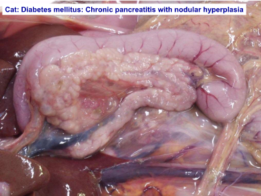
- Diabetes Mellitus
- Aetiology
- Relative or absolute lack of Insulin from pancreatic Beta cells (i.e. hypofunction)
- Destruction of islet cells secondary to pancreatitis
- The exocrine pancreas initially is inflamed, the rxn spreads to & involves the islets of Langerhans
- Condition can be chronic & relapsing, with gradual replacement of pancreatic tissue by fibrosis
- The exocrine pancreas initially is inflamed, the rxn spreads to & involves the islets of Langerhans
- Amyloid Deposition in Islets of Langerhan(Cats)
- Note: Older Cats often have scattered amyloid deposits in islets w/o signs of diabetes mellitus
- Idiopathic Pancreatic Atrophy
- Hypoplasia
- Destruction of islet cells secondary to pancreatitis
- Relative or absolute lack of Insulin from pancreatic Beta cells (i.e. hypofunction)
- Pathology
- Reduced availability of Insulin -> Hyperglycaemia
- Weight loss & weakness
- Reduced resistance to infection (Leukocyte function is impaired)
- Hepatic fatty change (can lead to cirrhosis in some cases)
- Cataracts -> bilateral lens opacity (due to sorbitol pathway metabolism of glucose by the lens)
- Renal glomerular sclerosis (Deposits of glycoprotein in the glomeruli)
- Cataracts -> bilateral lens opacity (due to sorbitol pathway metabolism of glucose by the lens)
- Hepatic fatty change (can lead to cirrhosis in some cases)
- Reduced resistance to infection (Leukocyte function is impaired)
- Weight loss & weakness
- Reduced availability of Insulin -> Hyperglycaemia
- Common in Dogs (esp. Females)
- Aetiology
- Endocrine Pancreatic Neoplasia
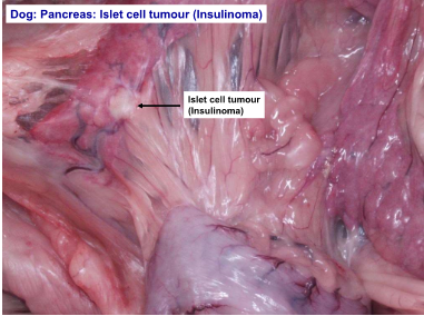
- Insulinoma
- Neoplasia of pancreatic Beta cells
- Carcinomas are more common than adenomas
- Usually functionally active -> Marked systemic effects
- Functional tumors => Hypoglycaemia
- Over time, hypoglycaemia => Neuronal necrosis
- More common in duodenal limb of pancreas
- Weakness, ataxia, altered temperament (which can progress to seizures & collapse)
- Functional tumors => Hypoglycaemia
- Usually functionally active -> Marked systemic effects
- Gastrinoma
- Uncommon compared to insulinoma
- Gastrin causes hypersecretion of gastric acid -> Ulceration of gastrointestinal mucosa
- Vomiting, diarrhoea & weight loss
- Invade pancreas, then spread to lymph nodes & liver
- Vomiting, diarrhoea & weight loss
- Glucagonoma
- Rare
- Glucagon induces hyperglycaemia
- Insulinoma
- Diabetes Mellitus
Thyroid Gland
- Develomental Disorders
- Accessory thyroid tissue
- Common
- Remnants of embryological development of thyroid gland
- Usually in mediastinum, often in heart base area
- Thyroglossal Duct Cysts
- In ventral cervical region, fluctuant cysts
- Can become neoplastic
- In ventral cervical region, fluctuant cysts
- Accessory thyroid tissue
- Hypothyroidism
- Imp. in Dog (Uncommon in other species)
- Idiopathic follicular atrophy
- Slowly progressive replacement of the gland by adipose tissue
- Lymphocytic thyroiditis
- Lymphocytic infiltration into the gland as a consequence of autoantibody formation
- Extrathyroidal Effects of Hypothyroidism
- Reduced BMR
- Skin
- Bilaterally symmetrical alopecia (esp. tail & neck area)
- Hyperkeratosis & Hyperpigmentation
- Myxoedema can lead to thickening of the skin (due to dermal accumulation of glycosaminoglycans & hyaluronic acid)
- Reproductive system
- Reduced sperm count, reduced/absent oestrus
- Metabolic alterations
- Marked increase in cholesterol levels, which in turn can lead to enlarged liver, atherosclerosis & glomerular & corneal lipidosis
- Hyperthyroidism
- Common in Cats
- Even w/ functional thyroid neoplasia in Dogs, it is uncommon to see hyperthyroidism since they are more able to excrete excess thyroid hormones
- Polyuria, Polydypsia, Polyphagia
- Weight loss
- Hyperexcitability
- Heat intolerance
- Cardiac alterations (Hypertrophic cardiomyopathy, Tachycardia, Dysrhythmia)
- Heat intolerance
- Hyperexcitability
- Weight loss
- Common in Cats
- Thyroid Gland Hyperplasia
- Goitre
- Non-neoplastic, non-inflammatory enlargement of the thyroid gland
- Diffuse goitre occurs in animals w/ an iodine deficient diet
- Nodular Hyperplasia
- Multiple small nodules within the gland
- Usually functional in the Cat (rarely functional in other species)
- Goitre
- Thyroid Gland Neoplasia
- Follicular adenoma
- Discrete capsule & usually solitary (compare to nodular hyperplasia)
- More common in Cats
- Follicular Carcinoma
- Locally invasive & often metastasize
- Can arise in extrathyroidal tissue
- More common in Dogs
- C-cell Neoplasia
- Most common in Bulls
- Thought to relate to high Ca2+ diet causing stimulation of the thyroid C-cells
- May be found in association with phaeochromocytomas & pituitary adenomas (multiple endocrine neoplasia - MEN)
- Most common in Bulls
- Follicular adenoma
- Develomental Disorders
Parathyroid Glands
- Hyperparathyroidism
- Primary Hyperparathyroidism
- Chief cell neoplasia
- Adenoma more common than carcinoma
- Most common in Dogs
- Often functional tumors (Increased levels of PTH) => Bone resorption & hypercalcaemia
- May cause pathological fractures of the bones
- Secondary Hyperparathyroidism
- Renal secondary hyperparathyroidism
- Nutritional secondary hyperparathyroidism (Low Ca2+ &/or high P)
- Pseudohyperparathyroidism
- Humoral hypercalcaemia of malignancy (e.g. apocrine adenocarcinoma of anal sac, lymphosarcoma, metastatic neoplasia to the bone)
- Primary Hyperparathyroidism
- Hypoparathyroidism
- Most common in small breed Dogs
- Usually caused by lymphocytic thyroiditis
- Clinical Signs (relate to consequences of progressive decline in serum Ca2+): Neuromuscular excitability
- Note: Potential to remove parathyroid glands during surgery
- Hyperparathyroidism
Chemoreceptor Organs
- Present at several sites (including carotid & aortic bodies) in many areas of the body -> Heart base tumors are most frequent
- Aortic body Adenoma & carcinoma
- Most common in Dogs (brachycephalic breeds)
- Adenomas are more common than carcinomas (most often seen as a mass of or multiple nodules around the base of the heart)
- Carcinomas can invade the pulmonary artery or atria (although distant metastases are uncommon)
- They are non-functional, but can cause problems due to their space occupying effect
- Carcinomas can invade the pulmonary artery or atria (although distant metastases are uncommon)
- In addition to aortic body tumors, ectopic thyroid tissue can cause tumor formation in the region of the base of the heart
- Carotid body adenoma & carcinoma
- Arise near bifurcation of common carotid artery
- Due to proximity to the artery and nerves -> excision/biopsy can be difficult
- Carotid body tumors tend to be more malignant then aortic body
- Due to proximity to the artery and nerves -> excision/biopsy can be difficult
- Arise near bifurcation of common carotid artery
- Multicentric tumors of chemoreceptor tissue occur (esp. in brachycephalic breeds)
Imagens e arquivos do mapa
Informações
- Descrição
- Doctorate Pathology (Systems Pathology) Mapa Mental sobre Endocrine Pathology, criado por melian.yates em 06-12-2013.
- Criado por
- melian.yates
- Visualizações
- 125
- Tags
- Avaliação