
Mindmap
Skin Path 1...
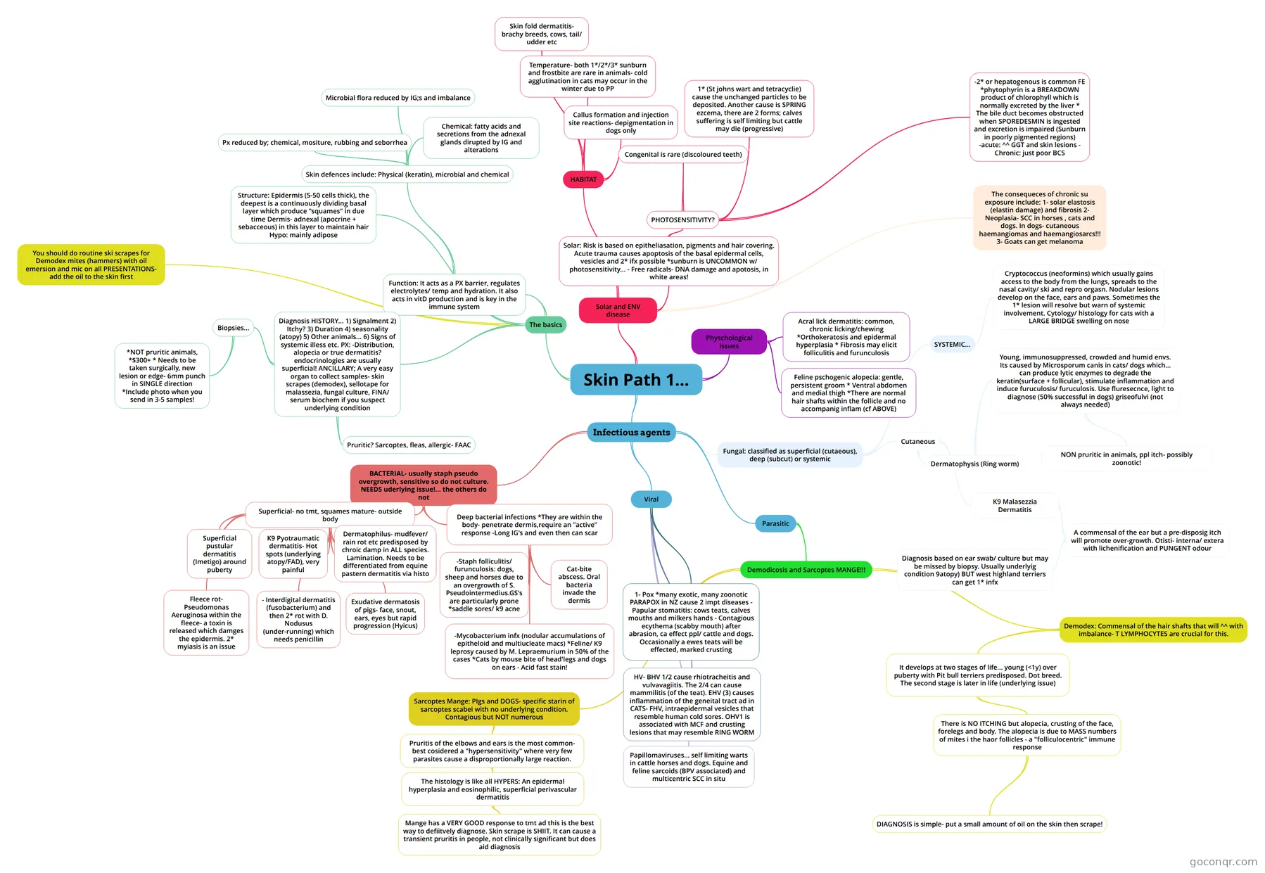
Mind map: Skin pathology
Inhalt der Mindmap
Skin Path 1...
The basics
- Function: It acts as a PX barrier, regulates electrolytes/ temp and hydration. It also acts in vitD production and is key in the immune system
- Skin defences include: Physical (keratin), microbial and chemical
- Px reduced by; chemical, mositure, rubbing and seborrhea
- Microbial flora reduced by IG;s and imbalance
- Chemical: fatty acids and secretions from the adnexal glands dirupted by IG and alterations
- Skin defences include: Physical (keratin), microbial and chemical
- Structure: Epidermis (5-50 cells thick), the deepest is a continuously dividing basal layer which produce "squames" in due time Dermis- adnexal (apocrine + sebacceous) in this layer to maintain hair Hypo: mainly adipose
- Diagnosis HISTORY... 1) Signalment 2) Itchy? 3) Duration 4) seasonality (atopy) 5) Other animals... 6) Signs of systemic illess etc. PX: -Distribution, alopecia or true dermatitis? endocrinologies are usually superficial! ANCILLARY; A very easy organ to collect samples- skin scrapes (demodex), sellotape for malassezia, fungal culture, FINA/ serum biochem if you suspect underlying condition
- Pruritic? Sarcoptes, fleas, allergic- FAAC
- Biopsies...
- *NOT pruritic animals, *$300+ * Needs to be taken surgically, new lesion or edge- 6mm punch in SINGLE direction *Include photo when you send in 3-5 samples!
- You should do routine ski scrapes for Demodex mites (hammers) with oil emersion and mic on all PRESENTATIONS- add the oil to the skin first
- Function: It acts as a PX barrier, regulates electrolytes/ temp and hydration. It also acts in vitD production and is key in the immune system
Solar and ENV disease
- Solar: Risk is based on epitheliasation, pigments and hair covering. Acute trauma causes apoptosis of the basal epidermal cells, vesicles and 2* ifx possible *sunburn is UNCOMMON w/ photosensitivity... - Free radicals- DNA damage and apotosis, in white areas!
- PHOTOSENSITIVITY?
- Congenital is rare (discoloured teeth)
- 1* (St johns wart and tetracyclie) cause the unchanged particles to be deposited. Another cause is SPRING ezcema, there are 2 forms; calves suffering is self limiting but cattle may die (progressive)
- -2* or hepatogenous is common FE *phytophyrin is a BREAKDOWN product of chlorophyll which is normally excreted by the liver * The bile duct becomes obstructed when SPOREDESMIN is ingested and excretion is impaired (Sunburn in poorly pigmented regions) -acute: ^^ GGT and skin lesions - Chronic: just poor BCS
- PHOTOSENSITIVITY?
- The consequeces of chronic su exposure include: 1- solar elastosis (elastin damage) and fibrosis 2- Neoplasia- SCC in horses , cats and dogs. In dogs- cutaneous haemangiomas and haemangiosarcs!!! 3- Goats can get melanoma
- HABITAT
- Callus formation and injection site reactions- depigmentation in dogs only
- Temperature- both 1*/2*/3* sunburn and frostbite are rare in animals- cold agglutination in cats may occur in the winter due to PP
- Skin fold dermatitis- brachy breeds, cows, tail/ udder etc
- Solar: Risk is based on epitheliasation, pigments and hair covering. Acute trauma causes apoptosis of the basal epidermal cells, vesicles and 2* ifx possible *sunburn is UNCOMMON w/ photosensitivity... - Free radicals- DNA damage and apotosis, in white areas!
Physchological issues
- Acral lick dermatitis: common, chronic licking/chewing *Orthokeratosis and epidermal hyperplasia * Fibrosis may elicit folliculitis and furunculosis
- Feline pschogenic alopecia: gentle, persistent groom * Ventral abdomen and medial thigh *There are normal hair shafts within the follicle and no accompanig inflam (cf ABOVE)
Infectious agents
- BACTERIAL- usually staph pseudo overgrowth, sensitive so do not culture. NEEDS uderlying issue!... the others do not
- Superficial- no tmt, squames mature- outside body
- Superficial pustular dermatitis (Imetigo) around puberty
- K9 Pyotraumatic dermatitis- Hot spots (underlying atopy/FAD), very painful
- Dermatophilus- mudfever/ rain rot etc predisposed by chroic damp in ALL species. Lamination. Needs to be differentiated from equine pastern dermatitis via histo
- Exudative dermatosis of pigs- face, snout, ears, eyes but rapid progression (Hyicus)
- - Interdigital dermatitis (fusobacterium) and then 2* rot with D. Nodusus (under-running) which needs penicillin
- Fleece rot- Pseudomonas Aeruginosa within the fleece- a toxin is released which damges the epidermis. 2* myiasis is an issue
- Deep bacterial infections *They are within the body- penetrate dermis,require an "active" response -Long IG's and even then can scar
- -Staph folliculitis/ furunculosis: dogs, sheep and horses due to an overgrowth of S. Pseudointermedius.GS's are particularly prone *saddle sores/ k9 acne
- Cat-bite abscess. Oral bacteria invade the dermis
- -Mycobacterium infx (nodular accumulations of epitheloid and multiucleate macs) *Feline/ K9 leprosy caused by M. Lepraemurium in 50% of the cases *Cats by mouse bite of head'legs and dogs on ears - Acid fast stain!
- Superficial- no tmt, squames mature- outside body
- Viral
- 1- Pox *many exotic, many zoonotic PARAPOX in NZ cause 2 impt diseases - Papular stomatitis: cows teats, calves mouths and milkers hands - Contagious ecythema (scabby mouth) after abrasion, ca effect ppl/ cattle and dogs. Occasionally a ewes teats will be effected, marked crusting
- HV- BHV 1/2 cause rhiotracheitis and vulvavagiitis. The 2/4 can cause mammilitis (of the teat). EHV (3) causes inflammation of the geneital tract ad in CATS- FHV, intraepidermal vesicles that resemble human cold sores. OHV1 is associated with MCF and crusting lesions that may resemble RING WORM
- Papillomaviruses... self limiting warts in cattle horses and dogs. Equine and feline sarcoids (BPV associated) and multicentric SCC in situ
- Parasitic
- Demodicosis and Sarcoptes MANGE!!!
- Demodex: Commensal of the hair shafts that will ^^ with imbalance- T LYMPHOCYTES are crucial for this.
- It develops at two stages of life... young (<1y) over puberty with Pit bull terriers predisposed. Dot breed. The second stage is later in life (underlying issue)
- There is NO ITCHING but alopecia, crusting of the face, forelegs and body. The alopecia is due to MASS numbers of mites i the haor follicles - a "folliculocentric" immune response
- DIAGNOSIS is simple- put a small amount of oil on the skin then scrape!
- There is NO ITCHING but alopecia, crusting of the face, forelegs and body. The alopecia is due to MASS numbers of mites i the haor follicles - a "folliculocentric" immune response
- It develops at two stages of life... young (<1y) over puberty with Pit bull terriers predisposed. Dot breed. The second stage is later in life (underlying issue)
- Sarcoptes Mange: PIgs and DOGS- specific starin of sarcoptes scabei with no underlying condition. Contagious but NOT numerous
- Pruritis of the elbows and ears is the most common- best cosidered a "hypersensitivity" where very few parasites cause a disproportionally large reaction.
- The histology is like all HYPERS: An epidermal hyperplasia and eosinophilic, superficial perivascular dermatitis
- Mange has a VERY GOOD response to tmt ad this is the best way to defiitvely diagnose. Skin scrape is SHIIT. It can cause a transient pruritis in people, not clinically significant but does aid diagnosis
- The histology is like all HYPERS: An epidermal hyperplasia and eosinophilic, superficial perivascular dermatitis
- Pruritis of the elbows and ears is the most common- best cosidered a "hypersensitivity" where very few parasites cause a disproportionally large reaction.
- Demodex: Commensal of the hair shafts that will ^^ with imbalance- T LYMPHOCYTES are crucial for this.
- Demodicosis and Sarcoptes MANGE!!!
- Fungal: classified as superficial (cutaeous), deep (subcut) or systemic
- Cutaneous
- Dermatophysis (Ring worm)
- Young, immunosuppressed, crowded and humid envs. Its caused by Microsporum canis in cats/ dogs which... can produce lytic enzymes to degrade the keratin(surface + follicular), stimulate inflammation and induce furuculosis/ furuculosis. Use fluresecnce, light to diagnose (50% successful in dogs) griseofulvi (not always needed)
- NON pruritic in animals, ppl itch- possibly zoonotic!
- Young, immunosuppressed, crowded and humid envs. Its caused by Microsporum canis in cats/ dogs which... can produce lytic enzymes to degrade the keratin(surface + follicular), stimulate inflammation and induce furuculosis/ furuculosis. Use fluresecnce, light to diagnose (50% successful in dogs) griseofulvi (not always needed)
- K9 Malasezzia Dermatitis
- A commensal of the ear but a pre-disposig itch will promote over-growth. Otisti- interna/ extera with lichenification and PUNGENT odour
- Diagnosis based on ear swab/ culture but may be missed by biopsy. Usually underlyig condition 9atopy) BUT west highland terriers can get 1* infx
- A commensal of the ear but a pre-disposig itch will promote over-growth. Otisti- interna/ extera with lichenification and PUNGENT odour
- Dermatophysis (Ring worm)
- SYSTEMIC...
- Cryptococcus (neoformins) which usually gains access to the body from the lungs, spreads to the nasal cavity/ ski and repro orgasn. Nodular lesions develop on the face, ears and paws. Sometimes the 1* lesion will resolve but warn of systemic involvement. Cytology/ histology for cats with a LARGE BRIDGE swelling on nose
- Cutaneous
- BACTERIAL- usually staph pseudo overgrowth, sensitive so do not culture. NEEDS uderlying issue!... the others do not
Informationen
- Beschreibung
- 3 Pathology Mindmap am Skin Path 1..., erstellt von rpbardoul am 03/09/2015.
- Erstellt von
- rpbardoul
- Aufrufe
- 1
- Bewertung