
Flashcards
Microbiology Test 3
Cards in this deck (142)
- 1Researchers converted skin cells into embryonic stem cells. They first inserted RNA complimentary for four embryonic genes into a virus, the resulting provirus inserted the genes into the skin cells' DNA. What virus can do this?Retroviruses
- 2General Characteristics of Viruses-Obligatory intracellular parasites
-Contain DNA or RNA (SS or DS; linear or circular)
-Contain protein coat called a capsid
-Some are enclosed by an envelope
-Some have spikes on envelope
-Most infect only specific types of cells in one host species
-Only seen by electron microscope
-Host range determined by specific host attachment sites and cellular factors - 3The 4 Main Viruses categories based on morphology (shape)1) Helical Virus
2) Polyhedral Virus
3) Enveloped Virus
4) Complex Virus - 4Comparing Viruses and bacteria: 1) Intracellular Parasites 2) Plasma Membrane 3) Binary Fission 4) Pass Through Bacteriological Filters1) Viruses: Yes; Bacteria: No
2) Viruses: No; Bacteria: Yes
3) Viruses: No; Bacteria: Yes
4) Viruses: Yes; Bacteria: No - 5Comparing VIruses and Bacteria 5) Possess both DNA and RNA 6) ATP-generating metabolism 7) Ribosomes 8) Sensitive to Antibiotics 9) Sensitive to interferon5) Viruses: No; Bacteria: Yes
6) Viruses: No; Bacteria: Yes
7) Viruses: No; Bacteria: Yes
8) Viruses: No; Bacteria: Yes
9) Viruses: Yes; Bacteria: No - 6What is one example of a helical virus?Ebola Virus
- 7What is a common polyhedral virus shape, and what are two examples of a polyhedral virus?Icosahedral (20-sided)
Poliovirus and Adenovirus - 8What is an example of an enveloped virus?Influenza virus
- 9What is an example of a complex virus?Orthopoxvirus
- 10What is a naked virus?A virus without an envelope
- 11Viral SpeciesA group of viruses sharing the same genetic information and ecological niche (host)
- 12Taxonomy of Viruses: What ending do we assign family names? What ending do we assign genus names?Family names end in -viridae
Genus names end in -virus - 13What is the family and genus of Human herpes virus (HHV-1 and HHV-2)Family: Herpesviridae
Genus: Herpesvirus - 14What is the family name and genus name of Human immunodeficiency virus (HIV-1 and HIV-2)Family: Retroviridae
Genus: Lentivirus - 15When growing viruses what factors must be considered?Viruses must be grown in living cells. Animal viruses may be grown in living animals or in embryonated eggs (usually grown on a membrane), while animal or plant viruses may be grown in cell cultures
- 16When bacteriophages form plaques on a lawn of bacteria, how many viruses does each plaque correspond to?One single virus. Can be expressed as plaque-forming units (PFU) that can be counted to determine the number of viruses
- 17Name four locations in an embryonated egg that a virus can be grown1) Chorioallantoic membrane inoculation
2) Amniotic inoculation
3) Allantoic inoculation
4) Yolk sac inoculation - 18What is a benefit of growing animal and plant viruses in a cell culture?Continuous cell lines may be maintained indefinitely
- 19What is the process of growing animal and plant viruses in cell cultures?1) A tissue is treated with enzymes to separate the cells
2) Cells are suspended in culture medium and usu grow in monolayer (stop growing when they touch each other)
3) Normal cells or primary cells grow in a monolayer across the glass/plastic container. Transformed cells or continuous cell cultures do not grow in a monolayer. Loss of contact inhibition tel you if it is infected (cytopathic effect). - 20Virus Identification Methods (3)1) Cytopathic Effect (CPE)
2) Serological Tests
3) Nucleic Acids - 21Virus Identification: Serological TestsDetect antibodies against viruses in a patient. Use antibodies to identify viruses in neutralization tests, viral hemagglutination, and Western Blot
- 22Virus Identification: Nucleic Acids-Southern Blotting
-Restriction Fragment Length Polymorphisms (RFLPs)
-PCR (polymerase chain reaction) - 23VirionsComplete virus infective units
- 24What are the different areas of a Viral One-step growth curveThe initial period has decreasing virions, leading into the eclipse period where there are no intact virions. The log growth phase is when virions are released from the host cell, and this peaks at the acute infection point. The log death phase occurs when there are no cells for the virus to use for reproduction
- 25Lytic CycleVirulent (lytic) phages. Eg T-even phages (T-2, T-4, t-6). The phage causes lysis and death of host cell
- 26Lysogenic cycleTemperate (lysogenic) phages. Eg bacteriophage lamda.
Prophage DNA is incorporated into host DNA. The host cell remains alive. Phage conversion: host cell exhibits new properties. Specialized transduction - 275 Steps of the Lytic Cycle1) Attachment (adsorption)
2) Penetration
3) Biosynthesis
4) Maturation
5) Release - 28The lytic cycle: Attachment (adsorption)Phage attaches by tail fibers to receptor sites on the bacterial cell wall
- 29The lytic cycle: PenetrationPhage lysozyme opens cell wall; tail sheath contracts to force tail core and DNA into cell (the only thing penetrating is the nucleic acid)
- 30The Lytic Cycle: BiosynthesisProduction of phage DNA and proteins (eclipse period). Like a virus factory
- 31The lytic cycle: MaturationAssembly of phage particles into complete virions
- 32The Lytic Cycle: ReleasePhage lysozyme breaks cell wall (lysis) and are released
- 33Multiplication of Animal Viruses: 1) AttachmentViruses attach to complementary attachment sites on the host cells plasma membrane
- 34Multiplication of Animal Viruses: 2) PenetrationBy pinocytosis (endocytosis) or fusion of envelope with plasma membrane. The entire virus gets taken in
- 35Multiplication of Animal Viruses: 3) UncoatingBy viral or host enzymes
- 36Multiplication of Animal Viruses: 4) BiosynthesisProduction of nucleic acid and proteins (differs for DNA vs. RNA viruses)
- 37Multiplication of Animal Viruses: 5) MaturationNucleic acid and capsid proteins assemble
- 38Multiplication of Animal Viruses: 6) ReleaseBy budding (enveloped viruses) or rupture
- 39Multiplication of DNA virusReplicate DNA In the nucleus of the host cell, using viral enzymes. Synthesize capsid in the cytoplasm using host cell enzymes. The virions are assembled w/in nucleus after capsid and other proteins migrate there. The virions are then transported along ER to the host cell's membrane for release
- 40Multiplication of RNA-containing VirusesAll occurs in the host cell's cytoplasm. These viruses have RNA-dependent RNA polymerase. Main differences lie in how mRNA and viral RNA are made.
- 41Bacteriophage viral multiplication (comparable slide for animal viruses)Attachment: tail fibers attach to cell wall proteins. Entry: viral DNA is injected into host cell. Uncoating is not required. Biosynthesis occurs in the cytoplasm. Then there is lysogeny, and then the host cell is lysed.
- 42Animal Virus Viral Multiplication (comparable slide for bacteriophages)Attachment sites are plasma membrane proteins and glycoproteins. Capsid enters by receptor-mediated endocytosis or fusion. Uncoating: enzymatic removal of capsid proteins. Biosynthesis in nucleus (DNA viruses) or cytoplasm (RNA viruses). Latency; slow viral infections; cancer. Enveloped viruses bud out of host cell; nonenveloped virsues rupture plasma membrane
- 43OncogenesActivated by chemicals, radiation, or viruses and transform normal cells into cancerous cells.
Viruses that turn on oncogenes are considered 'oncogenic' - 44Cancer: Transformed CellsHave increased growth, loss of contact inhibition, tumor-specific transplant antigens (on surface), or T antigens in nucleus. The genetic material of oncogenic viruses becomes integrated into the host cell's DNA (similar to lysogeny in bacteria). ~10% of cancers are caused by viruses
- 45Latent Viral InfectionVirus remains in asymptomatic in host cell for long periods. May reactivate due to changes in immunity: eg cold sores (Herpes simplex viruses), Shingles (adult chickenpox, or oncogenic viruses.
- 46Persistent chronic viral infectionsDisease processes occur over a long period; generally fatal. Subacute sclerosing panencephalitis (Measles virus); AIDS (HIV) is a classis example because slow increase in the number of virions over time.
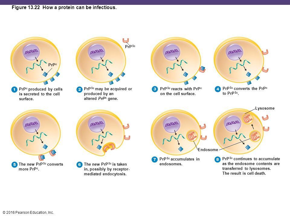
- 47PrionsProteinaceous infectious particle. Inhereted and transmissible by ingestion, transplant, and surgical instruments. PrP^c: Normal cellular prion protein, on cell surface
PrP^sc: Scrapie protein; accumulates in brain cells, forming plaques. Spongiform encephalopathies: sheep scrapie, CJD, and BSE - 48Plant VirusesEnter through wounds or via insects. Generally protected by an impermeable cell wall
- 49ViroidsInfectious naked RNA eg potato spindle tuber viroid (PSTV)
- 50How can a protein be infectious?
- 51A patient entered the hospital to have torn cartilage removed from her right knee. The surgery was scheduled as a same day procedure. Unfortunately, she subsequently developed pneumonia and wasn't released until 10 days later. How would you account for these events?The ventilation tube
- 52PathologyStudy of disease
- 53EtiologyStudy of the cause of disease
- 54PathogenesisThe development of disease
- 55InfectionColonization of the body by pathogens (+/- disease)
- 56DiseaseAn abnormal state in which the body is not functioning normally
- 57Normal microbiota (flora)permanently colonize the host and do not normally cause disease. Protect the host by occupying niches that pathogens might occupy, producing acids (eg human vagina pH = 3.5-4.5), and producing bacteriocins (inhibitory proteins)
- 58Transient microbiotaMay be present for days, weeks or months, then disappear.
- 59How many bacterial cells does the typical human body have?1 x 10^14
(compared to 1 x 10^13 human cells) - 60SymbiosisRelationship between normal microbiota and the host
- 61Commensalism definition and exampleOne organism benefits and the other is unaffected.
Eg. Staphylococcus epidermis on skin - 62Mutualism definition and exampleBoth organisms benefit. Eg E. coli bacteria in the large intestine
- 63Parasitism definition and exampleOne organism benefits at the expense of the other
Eg H1N1 virus particles - 64Microbial antagonism (competitive exclusion)A competition between normal and potentially pathogenic microbes
- 65What are bacteriocins?Inhibitory proteins
- 66ProbioticsLive microbes applied to or ingested into the body, intended to exert a beneficial effect.
- 67PrebioticsChemicals or substances that encourage growth of already present microbes
- 68Opportunistic pathogensPathogens that get where they shouldn't be. Eg E. coli in the bladder; causes cystitis
- 69Koch's postulates (Used to prove the cause of an infectious disease.)1) The same pathogen must be present in every case of the disease
2) Pathogen must be isolated from the diseased host and grown in pure culture
3) Pathogen from pure culture must culture disease when inoculated into healthy susceptible lab animal
4) Pathogen must be isolated from inoculated animal and shown to be original organism - 70Exceptions to Koch's postulatesSome pathogens can cause several disease conditions: eg Streptococcus pyogenes can cause sore throat, scarlet fever, skin infections, and osteomyelitis. Some pathogens ause disease only in humans, eg HIV. Some microbes have never been cultured Eg mycobacterium leprae (leprosy), Treponema pallidum (syphilis)
- 71SymptomChange in body function that is felt by a patient as a result of a disease (subjective - eg pain)
- 72SignA change in body that can be measured or observed as a result of disease (objective - eg temperature)
- 73SyndromeA specific group of signs and symptoms that accompany a disease
- 74Communicable diseaseA disease that is spread from one host to another, eg measles
- 75Contagious DiseaseA disease that is easily and rapidly spread from one host to another, eg measles
- 76Noncommunicable diseaseA disease that is not spread from one host to another, eg tetanus
- 77Sporadic diseaseDisease that occurs occasionally in a population; eg typhoid fever in Canada
- 78Endemic diseaseDisease that is constantly present in a population, eg common cold (rhinovirus)
- 79Epidemic diseaseDisease acquired by many hosts in a given area in a short time, eg influenza
- 80Pandemic diseaseWorldwide epidemic, eg AIDS
- 81Herd ImmunityImmunity to most of a population. If most of the population is protected (eg through vaccines) it minimizes transmission of that disease in that population)
- 82Acute vs chronic diseaseAcute: Symptoms develop rapidly but last a short time, eg influenza
Chronic: Symptoms develop slowly, eg TB - 83Subacute diseaseSymptoms between acute and chronic, eg subacute sclerosing panencephalitis
- 84Latent diseaseDisease with a period of no symptoms when the causative agent is inactive but then activates and produces symptoms. Eg shingles
- 85Local Infection vs Systemic InfectionLocal: Pathogens are limited to a small area of the body: eg abscesses
Systemic: An infection throughout the body, eg measles - 86Focal InfectionAn infection in a specific area of the body that results from a local infection that became systemic, eg from teeth or sinuses. Spreads and sets up shop elsewhere in the body
- 87Bacteremia Septicemia SepsisBacteremia: Bacteria in blood
Septicemia: Growth of bacteria in blood leading to sepsis
Sepsis: Toxic inflammatory condition arising from the spread of microbes, esp. bacteria or their toxins, from a focus of infection - 88Toxemia ViremiaToxemia: Toxins in the blood
Viremia: Viruses in the blood - 89Primary Infection Secondary InfectionPrimary: Acute infection that causes the initial illness
Secondary: Opportunistic infection after a primary (predisposing) infection eg Pneumocystic pneumonia with aids - 90Subclinical diseaseNo noticeable signs or symptoms (inapparent infection) eg carriers of hepatitis A virus. Can lie dormant.
- 91Predisposing Factors (8) that make the body more susceptible to disease1) Gender-short urethra in females
2) Inhereted traits - sickle cell gene
3) Climate and weather- incr colds in winter
4) Inadequate nutrition
5) Fatigue
6) Age
7) Lifestyle, occupation, habits (eg stress)
8) Chemotherapy - 92The stages of a diseaseIncubation period: No signs/symptoms
Prodromal Period: Mild signs/symptoms
Period of illness: Most severe signs/symptoms
Period of decline: signs and symptoms
Period of convalescence: May still be able to transmit disease to others - 93Reservoirs of InfectionContinual sources of infection
Humans: AIDS, gonorrhea, hepatitis.
Carriers may have inapparent infections or latent diseases
Animal: rabies, lyme disease
Nonliving: soil, eg botulism, tetanus; water, eg cholera, typhoid fever; food, eg trichinosis, salmonellosis - 94ZoonosesDiseases transmitted to humans from animals
- 95CarriersMay have inapparent infections or latent diseases (often asymptomatic)
- 963 Methods of Transmission of disease1) Contact
2) Vehicle Transmission
3) Vectors - 97Transmission of Disease: ContactDirect: Requires physical contact b/w infected and susceptible host (person-to-person)
Indirect: Spread by fomites (nonliving objects)
Droplet: Transmission via airborn droplets (<1m) - 98Transmission of Disease: Vehicle TransmissionTransmission by an inanimate reservoir: Eg water, food, air (>1m)
- 99Transmission of Disease: VectorsArthropods, esp. fleas, ticks, and mosquitoes. Transmit disease by 2 general methods:
Mechanical transmission: arthropod carries pathogen on feet (transport host)
and Biological transmission: Pathogen reproduces in vector (intermediate host); transmitted via bite or feces - 100Healthcare-Associated Infections (Nosocomial InfectionsAcquired by receiving treatment at a healthcare facility. Affect 5-15% of all hospital patients. 4th leading cause of death in Canada (8500-12000/yr). 30-50% are preventable through hand washing, gloves, masks, face shields, etc.
- 101What factors are required to acquired a Healthcare-Associated Infection?1) Microorganisms in healthcare associated environment
2) Compromised host
3) Chain of transmission (nurses, health care providers, etc) - 102Prevention and Control of Healthcare-Associated Infections (HAIs)'Break the chain of infection' by
-well staffed, well trained & stable teams
-more rigorous cleaning and proper supplies
-Stop and reverse contracting-out
-Reduce occupancy rates
-Mandatory standards, monitoring, and public reporting - 103PathogenicityThe ability to cause disease by overcoming the defenses of the host
- 104VirulenceThe degree of pathogenicity (how strong a pathogen is)
- 105Portals of EntryMucous membranes: gastrointestinal, genitourinary, and resp tracts; conjunctiva
Skin: hair follicles, sweat gland ducts
Parenteral route: beneath skin or mucous membranes eg punctures, injections, surgery
Many pathogens have preferred portals - 106Infectious dose 50 (ID50)Infectious dose for 50% of the test population
- 107Numbers of invading microbes for different portals of entry (ID50): 1) Skin 2) Inhalation 3) Ingestion1) 10-50 endospores
2) 10,000-20,000 endospores
3) 250,000-1,000,000 endospores - 108CapsulesPrevent phagocytosis by host cells. Phagocytosis can occur when antibodies produced against the capsule are present on the capsule surface (opsonisation). Examples: Sttreptococcus pneumoniae, Klebsiella pneumoniae, Haemophilus influenzae, and Bacillis anthracis
- 109Briefly explain opsonizationIt takes time for the host cell to form antibodies in response to the capsule. Once the capsule is coated with antibodies (on capsule surface), the phagocyte can consume the capsule
- 110Cell Wall Components (3)1) M protein
2) Opa protein
3) Mycolic acid - 111Cell wall component: M proteinOn cell surface and fimbriae resists phagocytosis and promotes adherence to surfaces. Eg Streptococcus pyogenes
- 112Cell Wall Component: Opa proteinOn outer membrane inhibits CD4+ lymphocytes (T helper cells) and allows attachment to host cells. Eg Neisseria gonorrhoeae
- 113Cell Wall Component: Mycolic acidWaxy lipid. Resists digestion by phagocytes. Eg Mycobacterium tuberculosis
- 114CoagulaseCoagulates (clots) fibrinogen in blood. Use coagulase test: find out good vs bad. Bad form clots (have coagulase).
Eg pathogenic Staphylococcus sp. - 115KinasesDigest fibrin clots. Eg streptokinase (fibrinolysin)
- 116HyaluronidaseHyrolyzses hyaluronic acid which cements connective tissue cells together
- 117CollagenaseHydrolyzes collagen of connective tissue (muscles, bones, tendons, etc)
- 118IgA proteasesDestroy IgA (secretory) antibodies that prevent adherence of pathogens to mucosa
- 119Antigenic variationAlter surface proteins to evade host immune system as antibodies are rendered ineffective. Can take place 1000x. An example is influenza virus.
- 120Penetration into the host cell cytoskeletonProduce surface invasins: Eg Salmonella alters host actin filaments to enter a host cell;
Use actin to move within cell and from one cell to the next: Eg Listeria, Shigella - 121How Pathogens Damage Host Cells: Using the Host Nutrients - IronBind directly to take in iron-transport proteins (lactoferrin, transferrin, ferritin) and hemoglobin of host. Secrete siderophores to strongly bind host's iron and steal it. Bacteria need iron to survive
- 122How Pathogens Damage Host Cells: Direct DamageDisrupt host cell function (use host for nutrients). Produce waste products. Lyse cell when released (viruses and intracellular bacteria). Toxins
- 123Toxin Toxigenicity ToxemiaToxin: Substance that contributes to pathogenicity
Toxigenicity: Ability of microbe to produce toxin
Toxemia: Presence of toxin in the host's blood - 124Intoxication ToxoidIntoxication: Presence of toxin without microbial growth
Toxoid: Inactivated exotoxin used in a vaccine to stimulate antitoxin production in body (preventative); eg tetanus toxoid. We make antibodies - 125AntitoxinAntibodies against a specific exotoxin (treatment). Eg tetanus antitoxin. We are given the antibodies.
- 1263 Main types of Exotoxins1) A-B Toxin (most common)
2) Membrane-Disrupting Toxins
3) Superantigens - 127ExotoxinsSource: Mostly gram +
Relation to microbe: By-products of growing cell
Chemistry: Protein
Fever?:No (most)
Neutralized by antitoxin?: Yes
LD50: Small (very lethal) - 128A-B ToxinA = active part
B = binding part (binds to host cell receptors)
Brought into cell by endocytosis.
Toxins causing botulism, tetanus, diphtheria, cholera, and anthrax - 129Membrane Distrupting Toxins (2 types)Lyse host's cells by
1) Making protein channels in the plasma membrane. Leukocidins kill phagocytic leukocytes (WBC's), Hemolysins destroy RBCs, Strepolysins destroy RBC's and WBC's
2) Disrupting phospholipid bylayer: Clostridial toxins causing food poisoning (C perfringens) & antibiotic-associated diarrhea (C difficile) - 130SuperantigensCause an intense immune response due to enormous release of cytokines from host T cells (overstimulate immune system). Symptoms: fever, nausea, vomiting, diarrhea, shock, and death. Eg. Staphylococcus aureus toxins causing food poisoning and toxic shock syndrome (TSS)
- 131EndotoxinsSource: Gram -ve only
Relation to microbe: Outer membrane
Chemistry: Lipid A
Fever?: Yes
Neutralized by antitoxin: No
LD50: relatively large (less lethal).
Heat stable (heat doesn't kill them) - 132When do we worry about endotoxins especially?When the outermembrane is destroyed and cytokines are released.
- 133Limulus amebocyte lysate (LAL) assayAmebocytes (WBCs) from horseshoe crab. Limulus Polyphemus, lyse in the presence of endotoxin. Clotting protein is released and resulting gel-clot is positive test for presence of endotoxin. Even if you kill all microbes, endotoxins still present.
- 134Viral Mechanisms for Evading Host Defences-Grow inside host cells -components of immune system can't reach them
-Hiding attachment sites from immune response (HIV binding sites on floors of valleys b/w surface folds on virus and hidden from antibodies but reachable by CD4 protein on T cells (WBCs) which virus binds to)
Visible effects of viral inf known as cytopathic effects (CPE) that may result in host cell death
CPE's vary with virus & used to diagnose many viral inf - 135Portals of Exit (5)Microbe generally uses same portal for entry and exit
1) Respiratory Tract
2) Gastrointestinal Tract
3) Genitourinary tract
4) Skin
5) Blood - 136Portals of Exit: Respiratory TractCoughing and sneezing
Eg influenza, TB, measles - 137Portals of Exit: Gastrointestinal TractFeces and saliva
Eg salmonellosis, shigellosis - 138Portals of Exit: Genitourinary TractUrine and vaginal secretions
Eg STD's - 139Portals of Exit: SkinDrainage from wounds
Eg herpes simlex, warts - 140Portals of Exit: BloodBiting arthropods (malaria) and needles or syringes (AIDS)
- 141Why is the number of invading microbes important regarding pathogenicity?The invading pathogen may overtake normal microbiota by sheer number
- 142—
Information
- Description
- Chapters 13-15
- Created by
- Shauna Ryner · Dec 2017
- Updated
- Mar 7, 2018
- Views
- 9
- Rating