
Mind map
Human Tissues
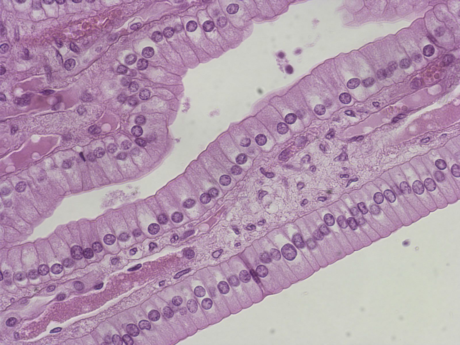
Map content
Human Tissues
Definition of Tissues: group of cells that have similar structure and that function together as a unit.
Four types of Tissues
- Muscular Tissues
- Epithelial Tissues
- General Characteristics
- Avascular, but innervated
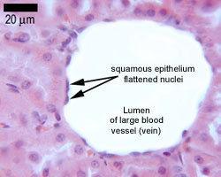
- Simple Squamous
- Function: absorption and filtration processes
- Location: blood and lymphatic vessels, air sacs of lungs, lining of the heart Function: secrets lubricating substance, allows diffusion and filtration
- pictures
- Location: blood and lymphatic vessels, air sacs of lungs, lining of the heart Function: secrets lubricating substance, allows diffusion and filtration
- Function: absorption and filtration processes
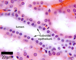
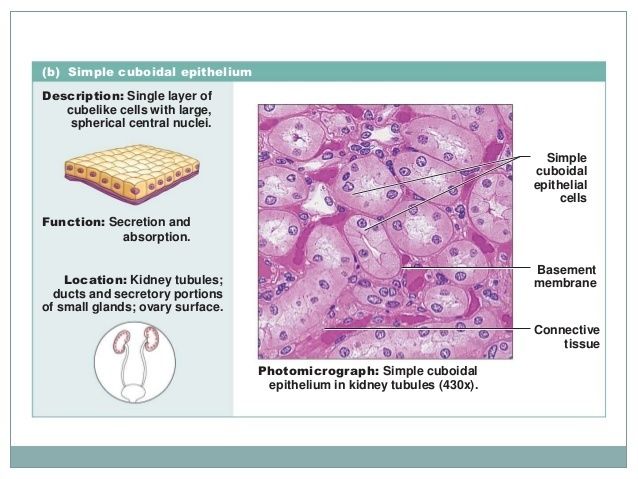
- Simple Cubical
- Function: absorption and filtration processes
- Location: secretory ducts of small glands, kidney tubules Function: allows secretion and absorbtion
- Function: absorption and filtration processes
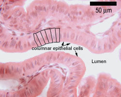
- simple columnar;
- Function: secretes acid, digestive enzymes and mucous.
- located in the stomach
- Function: secretes acid, digestive enzymes and mucous.
- protection, secretion, absorption, excretion, filtration, diffusion, and sensory reception.
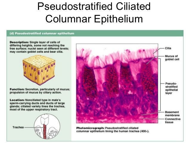
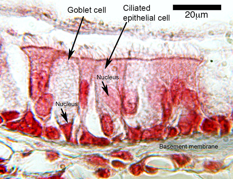
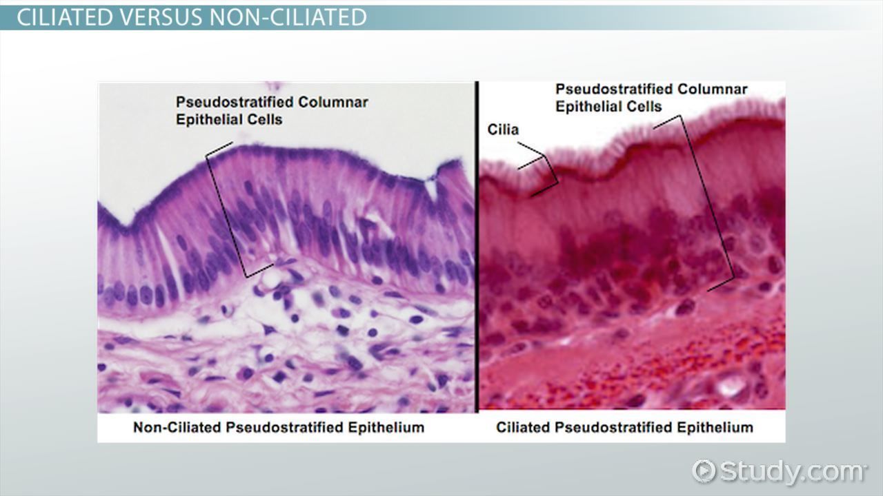
- pseudostratified
- Function: absorption and filtration processes
- Location: trachea and most of the upper respiratory tract (ciliated cells) Function: secretes mucus which is moved with cilia
- Function: absorption and filtration processes
- General Characteristics
- Nervous Tissue
- Connrctive tissue
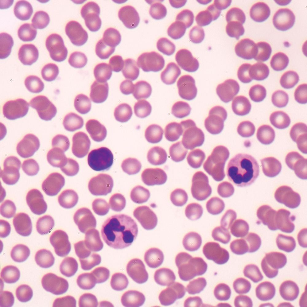
- blood
- location: throughout the body
- function: provides a mechanism by which gases, nutrients, wastes, and cells can be transported throughout the body.
- picture
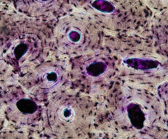
- bone
- location: in the skeleton
- Fucntion: supports body
- picture
- loose
- Areolar
- function: holds organs in place
- location: beneath the dermis layer and underneath epithelial tissue of all body system
- picture
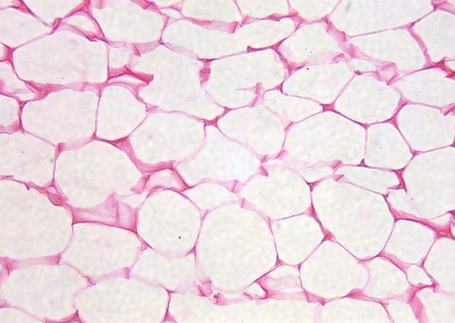
- Apidose
- location: beneath the skin, but is also found around internal organs.
- function: store energy in the form of fat, also cushions and insulates the body
- picture
- function: store energy in the form of fat, also cushions and insulates the body
- location: beneath the skin, but is also found around internal organs.
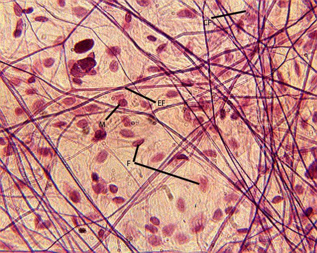
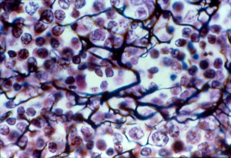
- Reticular
- Location: around the kidney, liver, the spleen, and lymph nodes, Peyer's patches as well as in bone marrow.
- function: support frameworks in the liver, lymphoid organs, capillary endothelial, and muscle fibers.
- picture
- function: support frameworks in the liver, lymphoid organs, capillary endothelial, and muscle fibers.
- Location: around the kidney, liver, the spleen, and lymph nodes, Peyer's patches as well as in bone marrow.
- Areolar
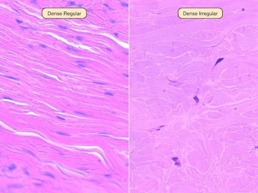
- dense
- regular
- location: in ligaments and tendons
- picture
- function: transfer forces to bones.
- location: in ligaments and tendons
- irregular
- location: in the reticular layer of the dermis and it’s also in the sclera and in the deeper skin layers
- picture:
- function: provides strength, making the skin resistant to tearing by stretching forces from different directions.
- location: in the reticular layer of the dermis and it’s also in the sclera and in the deeper skin layers
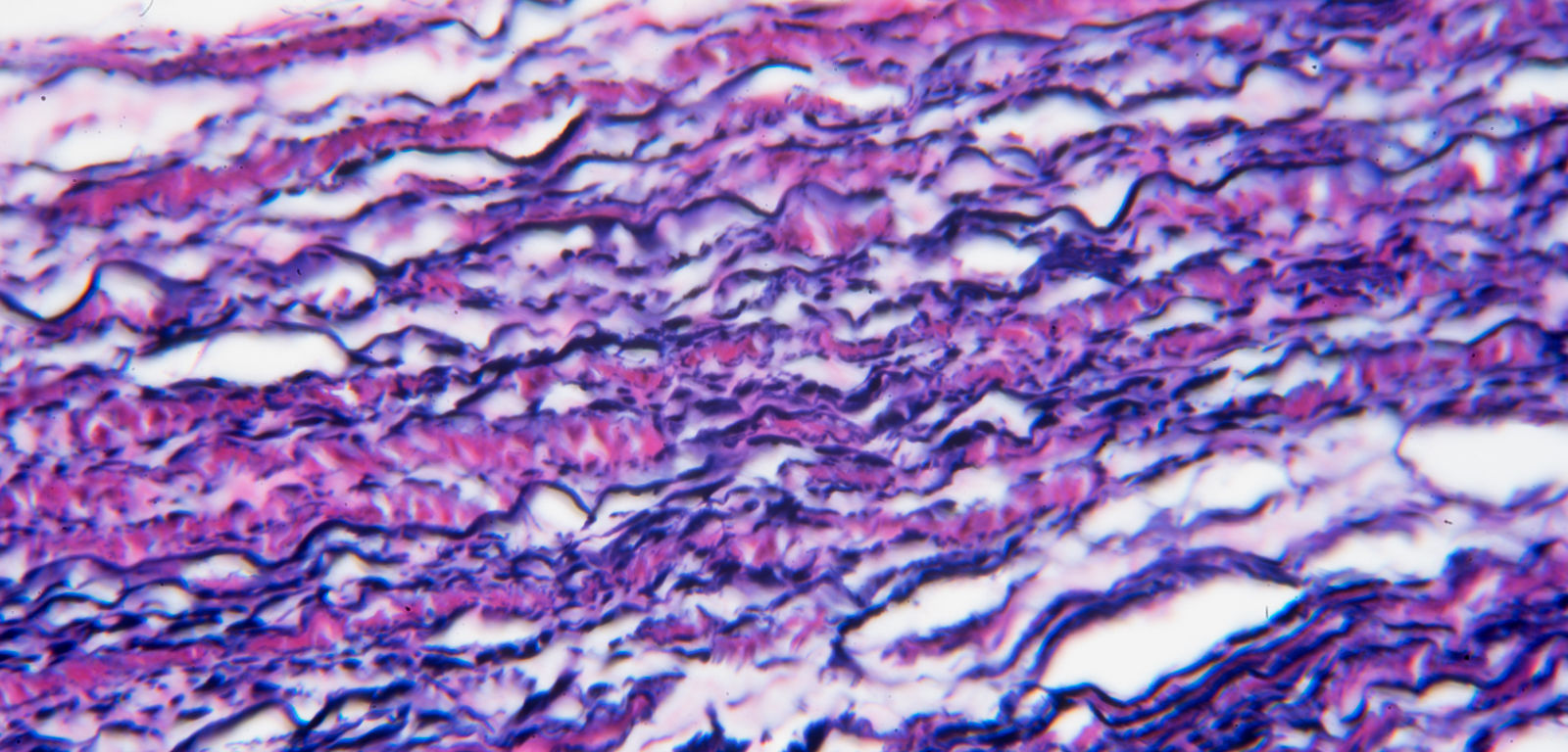
- Elastic
- location: tendons attach to bones, the intervertebral discs, and the pubic symphysis.
- picture
- function: hold connective tissues together, even during the movement of the body. It’s elastin is that after being stretched or compressed, it will return to its original shape.
- location: tendons attach to bones, the intervertebral discs, and the pubic symphysis.
- regular
- cartilage
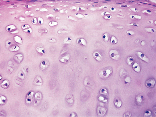
- hyaline
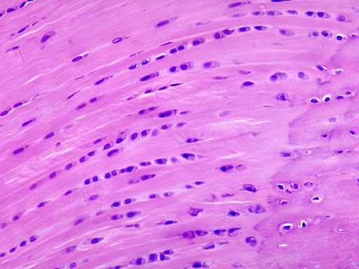
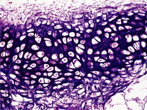
- location: in the nose, tracheal rings, and where the ribs join the sternum.
- Function: helps hold the body together and provides support and flexibility to different parts of the body.
- picture
- Function: helps hold the body together and provides support and flexibility to different parts of the body.
- location: in the nose, tracheal rings, and where the ribs join the sternum.
- Fibrocartilage
- location: located in regions where tendons attach to bones, the intervertebral discs, and the pubic symphysis.
- functions: helps fill in the torn part of the cartilage but not the ideal replacement.
- picture
- functions: helps fill in the torn part of the cartilage but not the ideal replacement.
- location: located in regions where tendons attach to bones, the intervertebral discs, and the pubic symphysis.
- Elastic
- location: located in the auricle of the ear and the epiglottis.
- function: provides strength, and elasticity, and maintains the shape of certain structures such as the external ear.
- picture
- hyaline
- blood
credita: https://www.histology.leeds.ac.uk/tissue_types/epithelia/epi_simple.php
nervous and muscle tissue
- sketal tissue
- location: It’s found in the muscles which are attached to the skeleton.
- fucntion: its function is to make up the muscle tissues connected to the skeleton and are important in locomotion.
- picture
- Cardiac Muscle
- location: The cardiac muscle is found in the walls of the heart.
- function: The function is to work to keep your heart pumping through involuntary movements.
- picture
- Smooth Muscle
- Location: The smooth muscle is located in the walls of hollow organs like your intestines and stomach.
- function: its function is to help contract your intestines to push the food through your body.
- picture
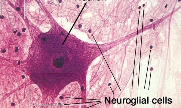
- Nervous Tissue
- location: it is found in the brain, spinal cord, and nerves.
- function: its function is to coordinate and control many body activities.
- picture
- sketal tissue
Images and files in the map
Information
- Description
- 12th grade human biology Mind Map on Human Tissues, created by Ashley Melgoza on 15/10/2020.
- Created by
- Ashley Melgoza
- Views
- 7
- History
-
- Created by Ashley Melgoza · Oct 7, 2020
- Copied by Ashley Melgoza · Oct 7, 2020
- Copied by Ashley Melgoza · Oct 15, 2020
- Rating