
Notes
TSEs
Content of the notes (1)
Page 1
Transmissible spongiform encephalopathies
In humans
Sporadic CJD
Occurs worldwide at an incidence of 1 /1000000
Familial disease
Familial CJG
Gerstmann-Straussler-Scheinker syndrome 1/10000000
Fatal familial insomnia
Atypical prion disease
Acquired disease
Kuru
Iatrogenic
vCJD
In Animals
Scrapie
Transmissible mink encephalopathy
Extinct
BSE origin
Chronic wasting disease (CWD) -US
Bovine spongiform encephalopathy (BSE)
Feline spongiform encephalopathy (FSE)
BSE origin
Other spongiform encephalopathies
Scrapie
Disease of sheep and goats
Characterised by
Ataxia
Tremors
Rubbing/scratching
Loss of condition
Death in 1-3 months
Endemic in UK
Incubation period of 2-5 yrs
Varied transmission
Adult animals can transmit it to other animals
Lamb exposed to prions in birth tissue shortly after birth.
Can also spread this way to other ewes
Inoculation
Agent can remain in environment (up to 50yrs)- don't know how
Progression to disease is under strong genetic control
Up to 15 genetic types of PrP gene with varied susceptibility scrapie
Genetic breeding programme
Varied strains
Bovine Spongiformform encephalopathy
Disease of cattle
Characterised by
Ataxia
Nervous or Aggressive behaviour
Loss of condition
Death in 1-6 months
Highly infectious
Can be spread to many other animals
Humans
Sheep
Goats
Cats
Epidemic in UK from 1985
Caused by contaminated feed
Incubation period of 5 years or more
Oral transmission (MBM) only
No transmission form cow to cow/ cow to calf
Little evidence of host genetic effect
Occurs as 2 different strains
MBM= meat and bonemeal
Chronic wasting disease (CWD)
First identified in captive deer in 1967 but then spread to wild deer
Found in
Free-ranging mule deer
White tailed deer
Rocky Mountain elk
Spread widely in North America + into Canada
Europe spread?
Found in Norway
Incubation period 16 months
Only TSE of wild populations
Only naturally spreading TSE
Limited genetic control os susceptibility
Unknown zoonotic potential
Hunting - dear to people??
variant Creutzfeldt-Jakob Disease
1996 UK government announced that vCJD had been detected in 10 people over 14 months
Clinical signs
Psychiatric
Depression
Anxiety
Delusions
Hallucinations
Neurological
Ataxia
Chorea
Myoclonus
Dementia
Pathology: Spongiform change and PrP-containind 'florid plaques' throughout the brain. In human prion diseases, plaques of this type are unique to vCJD
Mean duration of illness 14 months
Young age of onset: mean = 28 yrs (Range 12-74)
Prevalence of infection: 1 in 3000 (spleens)
Genetic control: All non-surgical human cases so far MM not MV or VV
Kuru
Background
Described in 1957 but may have started 1920
Shiver, trembling or laughing disease
Restricted to the Foré linguistic group in the highlands of New Guinea
Natural transmission
Foré ate their dead
Women and children ate internal organs and brains , men ate muscle
Cannibalism stopeed in 1957 encouraged by the governing Australian administration
Incidence of the disease declined from the 1960s, especially in cases of young children
Characteristics of TSE diseases
Long incubation period
Progressive and invariably fatal
Holes in brain give spongiform texture
No signs of fever
No signs of inflammation
No antibody response
No signs of a pathogen with a genome
No sign of infection
Causes
Partial purification of brain tissue of scrapie infected hamsters revealed that a certain protein was essential for infectivity
Procedures that destroy nucleic acid don't alter infectivity, unlike those that denature protein
Stanley prisoner suggested that infectious agents are proteinaceous: devoid of nucleic acid but capable of replication. He called them prions (proteinaceous infectious particles)
The prion protein is a host protein, encoded by the PrP (Protease-resistant protein) gene. It occurs in at least two form PrP^c is normal, PrP^Sc is infectious and associated with disease
PrP^c and PrP^Sc have an identical amino acid sequence but the differ in conformation
Prisoner suggested that PrP^Sc converts PrP^c to itself
PrP^Sc is partially resistant to digestion. In a normal cell PrP^c is constantly made and digested; PrP^Sc is not digested PrP^Sc is not digested and sos accumulates leading to disease
Can take years/decades
NB this is a hypothesis!
c=cellular
Sc=Scrapie
Uniqueness of prion diseases
Some prion diseases originate sporadically by mutation and are then transmissible
Pathogenesis
Oral route infection of sheep
Entry to body through the ileal Peyers patch
Dissemination through the lymphatic/vascular system to tissues of lymphoreticular system (LRS) including tonsil, spleen, reterophryngeal and mesenteric lymph nodes
Presence of PrP^Sc is detectable in the LRS weeks/months after infection, primarily within follicular dendritic cells (FDCs) of primary and secondary B cell follicles
Infection spreads from LRS through the ANS to the CNS
PrP^Sc accumulates primarily in glial cells and neurons. Within brain there is astrocytic gliosis, the accumulation of deposits of PrP^Sc, vacuolation and the characteristics of spongiform degeneration
Neuropathology
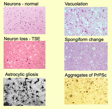
NB Astrocytic gliosis =aggregation of astrocytes
Diagnostics
Cant use PCR to detect nucleic acid or serology to detect antibody
Can use
Clinical disease
Histopathological detection of vacuolation , gliosis etc
Bioassay: especially in mice or hamsters
Expensive and time consuming
Molecular detection of PrP^Sc after treatment with proteinase K (PrPSc usually resists proteases)
No PK treatment PrP (c or Sc) deleted
PK treatment, no PrP detected = infer PrPc
PK treatment, PrP detected = infer PrPSc
Cyclic amplification of PrP^Sc from PrP^c
TSE strains
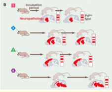
Specific scrapie isolates have characteristic effects which are consistently reproducible
Incubation period
Severity
Distribution
Brain pathology
Western blots are also distinct
The problem
Viral/Bacterial strain
Inoculate a host with strain X. Subsequently isolate strain X
Inoculate the same host with Y . Subsequently isolate strain Y
???
TSE strains
Inoculate a host with prion strain A. Subsequently isolate strain A
Inoculate the same host with strain B. Subsequently isolate strain B
???
Problems for the prion hypothesis
The same strain can be isolated from different host
Different host species have different PrP sequences - so strain can't be determined by the PrP amino acid sequence
The same host can be infected with different strains
A host only has one/two PrP types so strain cannot be determined by PrP amino acid sequence
Glycoforms
At least 8 prion strains have diff PrP^Sc conformations, suggesting that theological properties of prions are enciphered in conformation
Different strains also have unique combinations of glycoforms that are maintained upon transmission to mice
Species barrier
Transmission of agents limited by species barrier , characterised by extended incubation times which notable decrease as the agent is passaged though individuals of the recipient species
A large species barrier means a large infectious dose is needed for inoculum
Information
- Description
- Vet Med 1 (ID) Note on TSEs, created by Alice Butterworth on 02/02/2018.
- Created by
- Alice Butterworth · Feb 2018
- Updated
- Feb 2, 2018
- Views
- 8
- Rating