1. The Cytoskeleton
Slides in this set (43)
-
- Give an overview of the basic cytoskeletal components
- Explain basic principles governing microfilament assembly/disassembly
- Explain the role of actin cross-linking proteins in bundle/networks formation
- Explain how myosins interact with actin to act as motor proteins
- Explain the basic principles that govern the assembly and/or disassembly of intermediate filaments and microtubules
- Describe the function of intermediate filaments and microtubules
- How intermediate filaments and microtubules are organised within a cell
- Describe, with examples, how disrupted cytoskeleton can lead to disease conditions
-
Cytoskeleton: What it is and its functions
- What it is
- Highly ordered, dynamic network of filaments
- It is the dynamic nature that underpins its functions
- Functions
- Shape and motility of whole-cell and internal structures
- Critical for events such as cell morphology, migration, vesicle transport and cell division (cytokinesis and chromosome separation)
-
- Protein subunits self-assemble into polymers (filaments) -> form networks
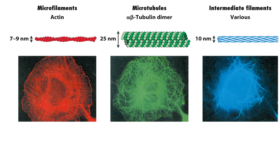
- 3 types of filaments and they form different networks with different functions
- Some overlap in networks= some cross-talk/cooperation between filaments
- Mictrotubules and int filaments have most overlap
- Microfilaments (7-9nm):
- Subunit= Actin
- Functions= Cellular movement, muscle contraction and cytokinesis (cell division)
- Microtubules(25nm):
- Subunit= Tubulin
- Functions= Scaffold to determine cell shape, provide transport tracks for organelles and vesicles to move along, form spindle fibres for separating chromosomes during mitosis
- Intermediate filaments(10nm):
- Subunit= Various intermediate filament proteins eg keratins, vimentin
- Function= Tensile cell strength
-
-
Actin drives eukaryotic cell movement
- Cell movement involves dramatic changes in cell shape -> driven by actin cytoskeleton
- Cell moves by putting out protrusions and pulling along surface
- Structure that facilitates cell migration:
- Actin bundles= extend from actin arc into leading edge (edge of cell in direction of movement)
- Stress fibres= extend from actin arc into cell tail
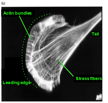
-
Actin drives eukaryotic cell movement
- The actin cytoskeleton underpins structures that facilitate cell movement:
- Protrusions:
- From leading edge, rich in actin that drive their extension
- Lamellipodium= Broad membrane extension that moves forward, typical of rapidly migrating cells
- Filopodia= Finer cytoplasmic extensions, typical of slower migrating cells
- Focal adhesions:
- Structures forming mechanical links between intracellular actin filaments and extracellular substrate
- Links filaments and extracellular matrix via integrins that span PM
- Assemble and dissassemble as cell moves
- Ruffles:
- Result from areas of leading edge not attached to substrate as cell moves
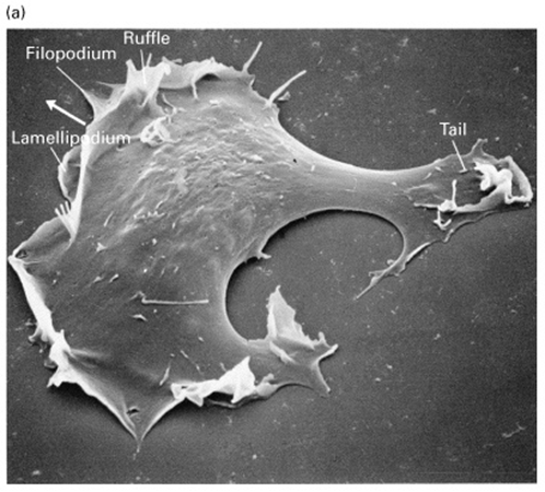
-
How activin generates force for movement
- Microfilaments generate force through assembly of G-actin into F-actin (Globular monomer to filamentous polymer)
- Features:
- End on end assembly of monomers
- F-actin adopts a tight helical structure
- Repeating unit (complete helical turn) = 14 subunits (37nm long, 7nm diameter)
- G-actin
- Ubiquitous in all eukaryotes and highly conserved
- Multiple isoforms= differ in sequence,function, tissue specificity but same general structure: Globular protein, 2 lobes separated by cleft, each lope has 2 domains and hinge between domains I and III
- Hinge allows 2 lobes to move relative to one another and form the nucleotide binding cleft (nucleotide= ADP or ATP)
- Nucleotide binding is required for G-actin stability
- ATP- or ADP-bound G-actin are identical structures except for subdomain 2 (important treadmilling)
- G-actin monomers have polarity (important for end on end stacking)
-
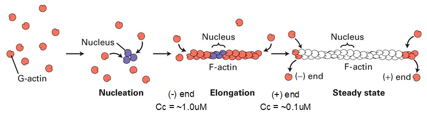
- Nucleation:
- 3 monomers form a trimer that serves as a nucleus
- Elongation:
- Monomers add to both ends in polarised manner (same orientation so F-actin has head and tail)
- Only add monomers when [G-actin] > critical concentration for polymerisation (Cc)
- [G-actin] increases, rate of assembly increases, filament depolymerises below Cc
-
-
F-actin polarity and treadmilling
- Polarity:
- Ends of F-actin have different Cc because of different structures of ATP- and ADP-bound actin
- Cc of assembly is always higher at - end than + end
- [G-actin] > 1uM= the filament extends in both directions but faster at + end
- 0.1uM< [G-actin] <1uM = deassembly at - end but assembly still at + end... treadmilling
- Treadmilling:
- Net loss from - and net gain at +
- Filament stays same length but subunits fall off - and add onto + -> movement of filament
- ADP-actin dissociates from - end
- ATP-actin polymerises at + end
-
- Activity necessary treadmilling:
- Once G-actin monomer joins to F-actin -> ATP is hydrolysed to ADP
- Hydrolysis of ATP is fairly slow so the longer the subunit has been apart of F-actin the more likely it is ADP-bound
- ADP- and ATP-bound G-actin have slightly different conformations -> ADP-actin has reduced affinity for neighbouring subunits, dissociates more readily
- ATP-actin has higher affinity so lower Cc
-
- Cycle: subunit added to + end moves towards - as more actin adds and hydrolyses to ADP-actin and dissociates at - end, once dissociated G-actin can exchanges ADP for ATP and repolymerise at +
-
More actin: actin in various architectures
- Actin filaments are organised into higher order architectures, collectively make up actin cytoskeleton
- Filopodium:
- Parallel bundles of actin filaments (all + at one end and - at other)
- Tightly packed actin microfilaments, finger-like projections
- Found in filopodia at leading edge
- Lamellipodium:
- Branched and cross-linked networks of filaments
- Cell cortex (gel-like network):
- Branched and cross-linked networks of filaments
- Structure underlies cell surface, provides tensile strength
- Stress fibres:
- Antiparallel bundles (each end has a mixture of + and -)
- Further apart than filopodium due to motor proteins inbetween
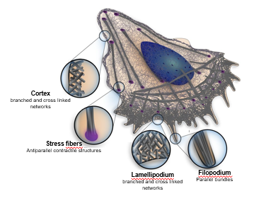
-
- Regulatory mechanisms to assemble and disassemble actin filaments include:
- Inhibition of spontaneous polymerisation
- Nucleation of new actin filaments
- Control of actin filament length
- Elongation/shortening of pre-existing actin filaments
- These and other functions are carried out by actin-binding proteins, which do 2 things:
- Regulate actin polymerisation and organisation
- Associate with monomers or already assembled actin filaments
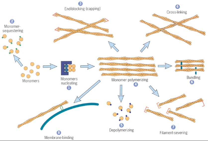
- Caption: Functions of actin-binding proteins
-
- Regulate polymerisation of G-actin
- Thymosin B4:
- Small peptide found in metazoan cells
- Binds tightly to ADP-G-actin (sequesters)
- Cant exchange nuc, no polymerisation at +
- Shortening of filaments
- Profilin:
- Present in most eukaryotes
- Binds ADP-G-actin opposite to nucleotide binding cleft
- Increases rate of ADP to ATP exchange due to where it binds
- Blocks addition of actin at - but not +, F-actin extends preferentially at + end
- Stops treadmilling - elongates filament
- Regulate nucleation of G-actin
- Formins:
- Provide template to bring 3 monomers of G-actin together to form nucleus
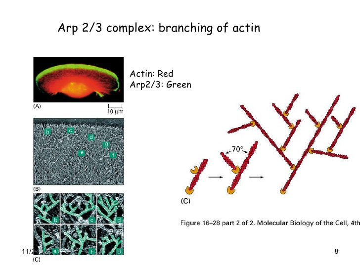
- Arp2/3:
- Consists of 2 Actin Related Proteins (+ 5 other subunits)
- Similarly forms nucleus which initiates branching of microfilaments: binding to a filament and nucleating to initiate polymerisation of new branch
-
-
- Two broad categories: severing and capping proteins and cross-linking proteins
- Severing and capping: regulate filament length
- Severing= break actin filaments into shorter fragments
- Capping= capping at 1 end: growth or shrinkage, at both ends: stabilised and loss of dynamic property
- Gelsolin: Capping (+) end and severing
- Dissolves mesh of actin filaments: binds to F-actin and severs filament, caps + end to prevent further growth, uncapped - end disassembles as usual
- Stimulus= high Ca2+ levels
- Inhibited by PIP2
- Cofilin(ADF= actin depolymerising factor): severing
- Binds ADP-actin: binding at - end of F increases depolymerisation of ADP-actin, binding deeper in filament causes severing, binding G-actin prevents nuc exchange (inhibits polymerisation at +
- Opposite to profilin (pro promotes assembly, cof promotes disassembly
-
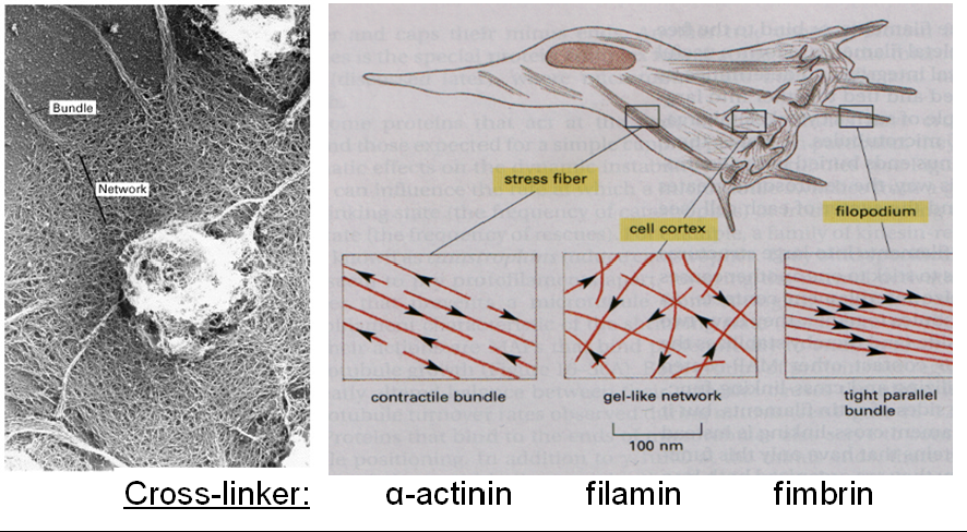
- Cross-linker proteins: organise filaments into networks and bundles (higher order actin architectures
- Cell cortex: coordinated by filamin
- Filopodium: coordinated by fimbrin
- Stress-fibres: coordinated by alpha-actinin (a-actinin)
- Cross-linkers share a common feature: actin binding domains
-
- Fimbrin
- Monomeric protein with 2 actin binding domains
- Each domain binds a F-actin filament
- Gives rise to tight parallel bundles
- a-Actinin
- One binding domain but forms a dimer (binding domain on either end of dimer)
- Gives rise to antiparallel bundles
- Filamin
- One binding domain but forms a dimer (binding domains at points of a V)
- Bind and organises filaments into networks
-
- Specialised cross-linkers:
- Spectrin:
- Tetrameric: 2 alpha and 2 beta chains, 2 binding domains (1 binding domain per aB chain, so 1 at either end of tetramer)
- Forms meshwork that gives RBCs their shape (biconcave)
- Biconcave= high surface: volume to exchange oxygen to surrounding tissues
- Mutations cause loss of biconcave shape, cant exchange oxygen well, associated with anaemia
- Dystrophin
- Monomeric, part of a multi-protein complex connecting muscle fibre cytoskeleton (microfilaments) to ECM
- Single actin binding domain
- Important role in force generation, mutations underlie muscular dystrophies
-
Actin filaments as tracks for transport
- Actin cytoskeleton relies on motor proteins to play a role in intracellular transport
- Myosins
- Motor protein that carries transport vesicles along actin filaments
- Multi-gene family: different myosins have different functions
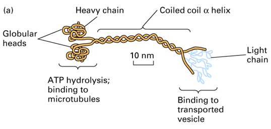
- Made of 1 or 2 heavy chains and some light (regulatory) chains
- Myosin I= monomer (1 heavy), myosin II and V= dimers (2 heavy)
- Bind actin through myosin "head" that has ATPase activity to generate force to "walk" along filaments
-
Actin filaments as tracks for transport
- Myosin I (monomer)
- Function: cytoskeleton-membrane interaction, links PM with F-actin filaments in filopodia and microvilli
- Myosin II (dimer)
- Function: contractile force for muscle contractions and cytokineses (tightens contractile ring between daughter cells)
- Myosin V (dimer)
- Function: cytoskeleton-membrane interaction, links F-actin filaments membrane of transport vesicles for power transport of vesicles
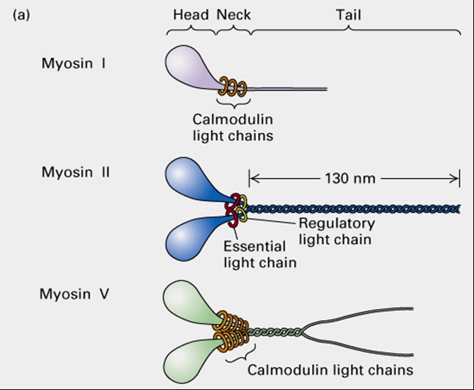
- Caption: myosin head and neck region conserved so vary in other structural components
-
Myosin II: generating contractile force
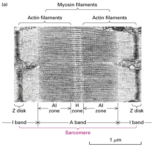
- In striated muscle, myosin II dimers are joined together so tails pack tightly to form thick filament, heads protrude out of tail at either end
- Sarcomere
- Thick filaments are interspersed within actin thin filaments, functional contractile unit of muscle
- Adjacent sarcomeres joined through Z disks which anchor actin filaments at end of sarcomeres
- Sliding filament model
- Muscle contracts: thick and thin filaments slide past each other, sarcomere shortens
-
Myosin II: generating contractile force
- Sarcomeres -> myofibrils-> myofibers-> muscle
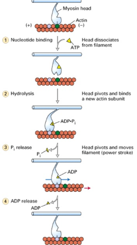
- Muscle contraction mechanism
- Sarcomere resting state= myosin head bound to F-actin via actin binding domain
- ATP binding causes conformational change in myosin (heavy chain), head dissociates
- ATP hydrolysis causes change in neck (hydrolysis products are trapped), head pivots back and re-binds filament further back
- Upon release of products, head swings back to normal position whilst bound to actin, provides power to slide actin and myosin past eachother, contract sarcomere (power stroke)
-
- Features of intermediate filaments:
- Large diversity in seq and size in contrast to microfilaments and microtubules
- 5 major classes of int filament proteins (subunits): Type I-V
- Main function: provide high tensile strength to buffer against external forces
- Expression of proteins is well characterised and immunostaining of some can be used to identify origin of metastatic tumours
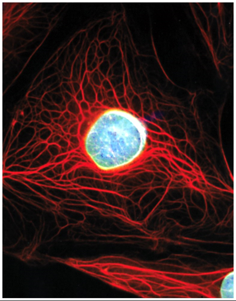
- Blue lamin staining- lamins make up nuclear int filaments, maintain nuclear str
- Red keratin staining- keratins make up cytosolic int filaments that extend into cell mem in skin epithelial cells (keratinocytes)
-
Intermediate filament proteins
- Keratins:
- 2 of 5 major classes: Type I= acidic keratins, Type II= basic keratins
- Found in outer epithelia
- Fibrous protein, prominent in skin, hair and nails
- Forms heterodimers of basic and acidic subunits of intermediate filaments, tight association between subunits gives strong structure
- Mutations associated with skin disease
- Vimentin:
- Type III intermediate protein
- Widely distributed: stromal tissues, lymphocytes, endothelial cells, fibroblasts
- Supports cell membranes and keeps nucleus/organelles in position (highly expressed in stromal tissues)
- Immunostaining
-
Intermediate filament proteins
- Neuronal intermediate filament proteins:
- Type IV
- Neurofilaments
- Structural roles in axons: determine and maintain axon diameter which controls speed of conduction of nerve impulses
- Parallel bundle arrangement of filaments within axons
- Lamins:
- Type V
- Forms fibrous network supporting inner nuclear membrane (expression of lamins restricted to nucleus)
- Also role in organising different types of chromatin and contribute to regulation of gene expression
- Immunostaining
-
Assembly of intermediate filaments
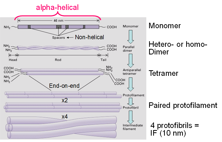
- General structure of intermediate filament proteins (monomer):
- Globular head(N-): role in assembly
- Globular tail(C-): dispensable for assembly, role in filament stability (facilitates lateral interactions within a filament)
- Both head and tail facilitate interactions between filaments and cellular components
- Extended alpha helical region that separates head and tail, backbone of filament
- Most sequence diversity in N- and C-, so they were thought to not have role in assembly (highly conserved) but truncation of these domains -> filament doesn't form properly
- Alpha helical core highly conserved
- Assembly is spontaneous- no ATP or GTP needed
-
Assembly of intermediate filaments
- Intermediate filaments wrap around each other -> parallel dimer
- 2 dimers go head to tail -> antiparallel tetramer
- Tetramers stack end-on-end -> protofilament
- 4 protofilaments wrap around -> 10nm diameter int filament
-
Int filaments: hetero- or homo-polymers
- Spacer sequences :
- Sequences within alpha-helical core determine which proteins can heteropolymerise: spacer sequences of different IF proteins need to be same length
- Keratins: heteropolymerise, always contain type I and type II keratins
- Type III filament proteins eg vimetins: homo or hetero with other IF proteins
-
Int filaments maintain cell integrity
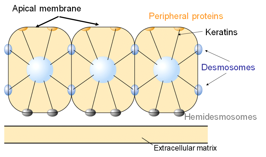
- Roles: anchor to cell junctions, maintain cell integrity and position of structures within cells:
- Keratin has a role in nuclear positioning- links nucleus to cell adherence molecules at PM
-
- Red lines= basement membrane that separates dermal and epidermal layers
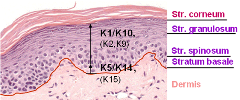
- Basal dermal layer: skin stem cells differentiate as they move up dermis, through differentiation expression patterns of keratins change:
- Basal keratinpcytes- express K5 and K14, allow tight association to basal membrane (K5 and 14 form parallel heterodimer in assembly)
- Progression to outer layers, lose 5 and 14 and gain expression of K1 and K10, allow more flexibility
-
Disease associated with int filaments
- Keratins: skin disease
- Mutations in K5 and K14. N-/C- mutations
- Cells at base of epidermis are weakened, cant properly adhere to basal membrane
- Epidermis and dermis easily separated -> weak skin easily wounded, blistering disease (EBS)
- Lamins: Hutchinson-Gilford progeria syndrome
- Rare premature aging
- Mutation in LMNA gene, abnormal form of nuclear int filament Lamin A
- Nuclear envelope becomes unstable and DNA is prone to damage
- Also associated with phenotypes: hair loss, cardiovascular disorders, diabetes, muscle atropy
-
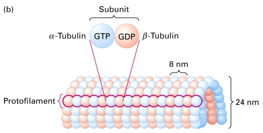
- Polymers of globular tubulins arranges into a tube (24nm)- 2X width of IFs, 3X MFs
- Subunit= heterodimers of one alpha and beta globular tubulin
- Dimers polymerise head to tail -> polarity (similar to MFs)
- Act as cellular tracks used by motor proteins eg kinesin and dyenin for transport of vesicles/organelles and separation of chromatids in mitosis
-
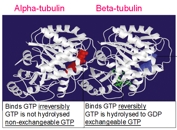
- Alpha-tubulin: binds GTP irreversibly, GTP not hydrolysed and not exchangeable
- Beta-tubulin: binds GTP reversibly, when bound hydrolysed to GDP then exchanged for GTP, hydrolysis and exchange occurs at microtubule ends (similar to MF assembly)
-
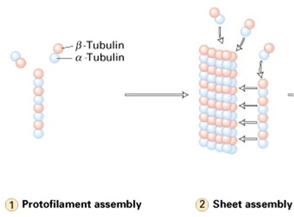
- Individual heterodimers assemble end-on-end in cytoplasm -> protofilament
- Several protofilaments associate laterally-> sheet (increase stability)
- Also simultaneously extends longitudinally through addition of more dimers onto sheet
- Sheet curves into hollow tube
-
Microtubules assembly: elongation
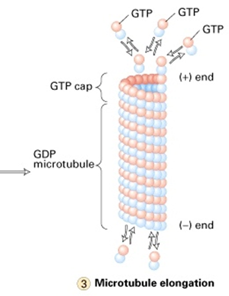
- Dimers can be added onto both ends( like in MFs): growth faster at +
- Beta(+ end), alpha(-end)
- GTP on beta is hydrolysed when subunit is part of tubule
- Closer dimer is to + end more likely have GDP (hydrolysed GTP)
- Rate of addition of dimers at + > rate of GTP hydrolysis by beta-tubulin = cap formation at +
- Characteristic of rapidly growing tubule
- Stabilises tubule since GTP on beta-tub have higher affinity for neighbours than GDP
-
- Assembly is conc dependent (Cc):
- Dynamic instability= growth/shrinkage either side of Cc
- Dimer conc > Cc= polymerisation and growth (preferentially at +)
- As MT grows, pool of dimers depletes
- Dimer conc< Cc= MT disassembles (preferentially at -)
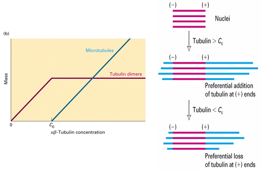
-
Microtubule organising centre (MTOC)
- MT in a cell are anchored at their - end onto the MTOC : located near nucleus, directs assembly and orientation of MTs, direction of vesicle traffic and orientation of organelles
- Animal cell MTOC= centrosome:
- Pair of centrioles made of short MTs that are linked through proteins in pericentriolar matrix
- Anchor - ends and leave + ends to grow/shrink
- During mitosis centrioles replicate and move to opposite poles, centrioles link MTs to sister chromatids and pull sister chromatids to opposite poles (using force they generate, dissassembly increases by 10 fold)
- Coordination- then actin ring will form and cytokinesis occurs
-
Microtubule organising centre (MTOC)
- Another MTOC= Basal body in flagella
- Cylindrical structure that sits at junction of flagellum
- Organises MTs, allowing to extend into flagella and create force for movement
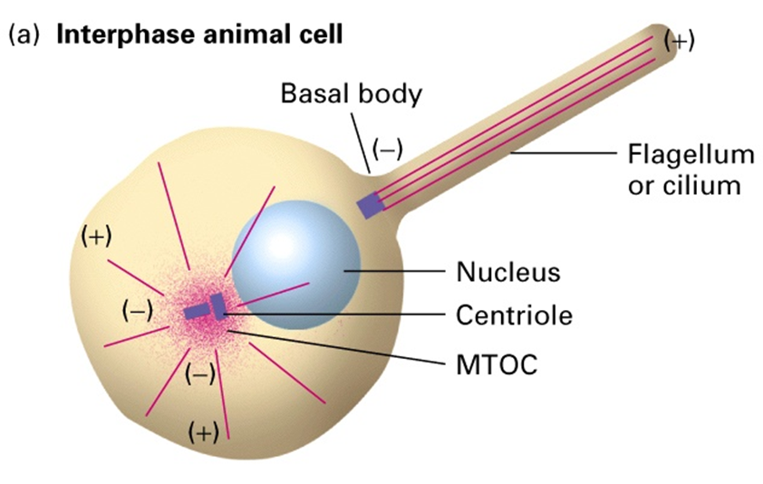
-
- Eg nerve cells with long dendrites that receive nerve impulses from other neurons have stable MTs:
- Consequence of MTOC= MTs all orientated in same direction
- In dendrites MTs show mixed polarity important to transport in both directions
- Axon regions are large distances so need to transport in both directions between cell body to nerve ending
-
Microtubules as transport tracks
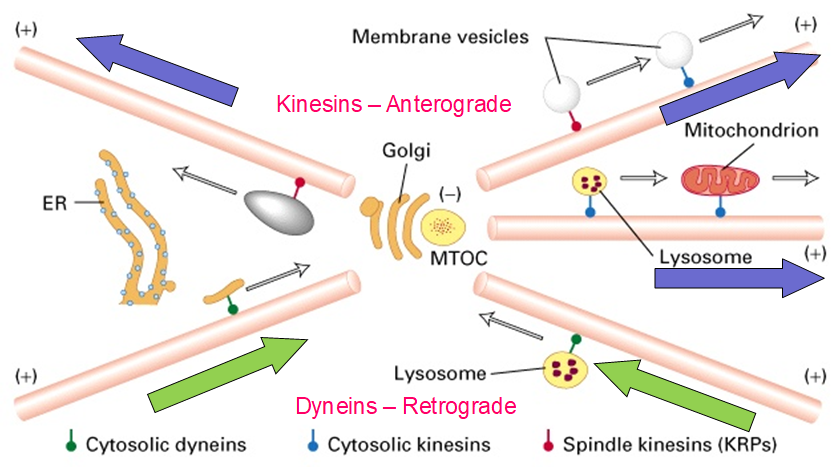
- Cellular tracks for trafficking vesicles, organelles and chromosomes inside a cell
- Used by MT motor proteins: kinesin and dynein that work in opposite directions to each other
- Kinesins: carry cargo away from MTOC from - to +
- Dyneins: carry cargo towards MTOC + to -
-
Microtubules as transport tracks
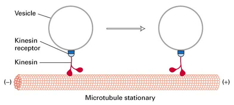
- Kinesin head domain (ATPase activity) docks with microtubules
- MT remains stationary (track) and vesicle is transported from - to +
- Walks along
-
Microtubules as transport tracks
- Regions within a cell where the vesicle travels may be MT-poor and MF-rich
- But single vesicles can have both kinesin (MT ) and myosin (MF) motors:
- Kinesin to get through cell body, move along MTs
- Myosin to get through cell cortex (less MTs, more MF), move along MTs
- Kinesin structural similarity to myosin :
- Forms dimer
- Have 2 head regions that bind to cytoskeleton
- Tail to bind to transport vesicle/mitochondria