
Flashcards
Biology AQA 3.1.3 Cells
Cards in this deck (14)
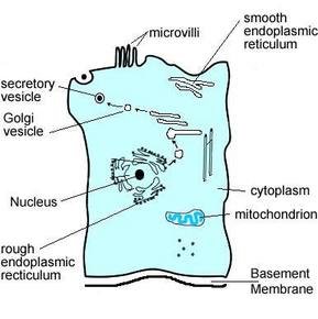
- 1Structure of an epithelial cell from the small intestine.
- 2—PLASMA MEMBRANE:
Appearance: found on the surface of animals cells and inside the wall of plant cells and prokaryotic cells. It is made up of proteins and lipids.
Function: regulates movement of substances in and out of cells. it also has receptors which responds to hormones and chemicals.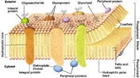
- 3—MICROVILLI:
Appearance: These are folds in the plasma membrane
Function: Involved in processes like absorption and they increase the surface area of the plasma membrane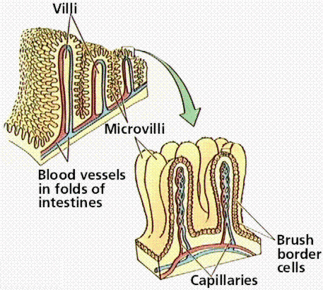
- 4—NUCLEUS:
appearance: A large organelle surrounded by a nuclear envelope( double membrane) It contains many pores and the nucleus contains chromatin and often a nucleolus
Function: Chormatin is made from proteins and this makes up DNA which controls the cells activities. The pores allow substances to move in and out and the nucleus makes ribosomes.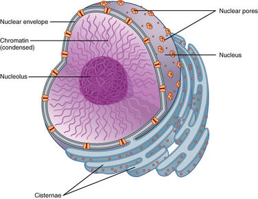
- 5—LYSOSOME:
Appearance: A round organelle surrounded by a membrane with no clear internal structure.
Function: Contains digestive enzymes and are used during phagocytosis and they digest invading cells or break down worn components of the cell.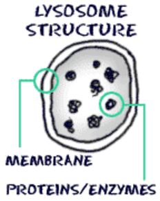
- 6—RIBOSOME:
Appearance: A small organelle that floats free or is attached to a rough endoplasmic reticulum.
Function: This is the site where proteins are made.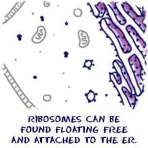
- 7—ENDOPLASMIC RETICULUM:
Appearance: SMOOTH: system of membranes enclosing a fluid filled space
ROUGH:Is similar to smooth but is covered in ribosomes.
Function: SEM synthesises and processes lipids. REM folds and processes proteins that have been made by ribosomes.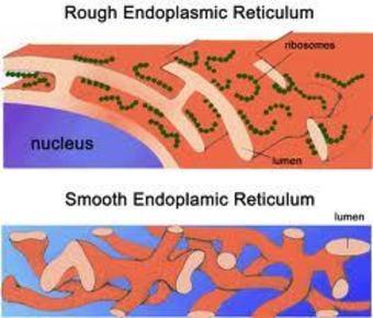
- 8—GOLGI APPARATUS;
Appearance: A group of fluid filled flattened sacs.
Function: Processes and packages new lips and proteins. It also makes lysosomes.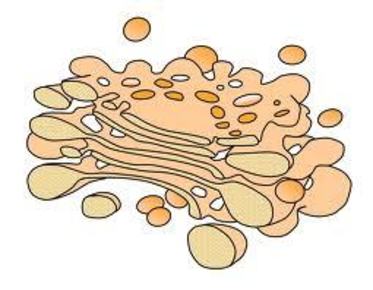
- 9—MITOCHONDRION:
Appearance: They have a double membrane and the inner membrane is folded to form cristae, inside this is the matrix which contains enzymes involved in respiration.
Function: The site of aerobic respiration. Found in large numbers are active and require lots of energy.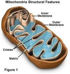
- 10Prokaryote: Before nucleusEukaryote: After Nucleus
- 11Transmission Electron Microscopes Electrons are fired onto the specimen which then absorbs electrons. this then causes an image to be produced on a screen *The whole thing must be in a vacuum *A complex staining process *Image is black and white * Specimen must be thing *Image might contain artefactsScanning Electron Microscopes
Same limitations as a transmission. However the specimen doesnt need to be thin as a 3D image can be produced. - 12Magnification: Magnification= Size of image/ size of object M= I/o (MILO)Resolution:
The minimum distanceof two objects for them to appear seperate - 13Cell Fractionation: the process where cells are broken up in a c cold, isotonic and buffered solution.Cold: Reduce enzyme activity
Isotonic: To prevent organelles bursting or shrinking
Buffered: to maintain a constant PH - 14Ultracentrifugation: Process by which fragments are seperated.1. The tube is spun at a slow speed.
2. The heaviest organelles (the nucleus) are forced to the bottom of the tubes, they then form a sediment.
3. The fluid at the top is removed leaving the nucleus
4. the fluid is then spun again and the heaviest sinks and the organelles continue to be seperated
Information
- Description
- Unit 1 Biology, Aqa specification 3.1.3
- Created by
- evie.daines
- Views
- 4,045
- Tags
- Rating